03.09.2020

The brand new  Levenhuk N38 Prepared Microscope Slides Set includes two kits of experiment slides — Levenhuk N18 and Levenhuk N20.

First, we will discuss the  Levenhuk N18 Prepared Microscope Slides Set. It includes slides for botanical and zoological studies.

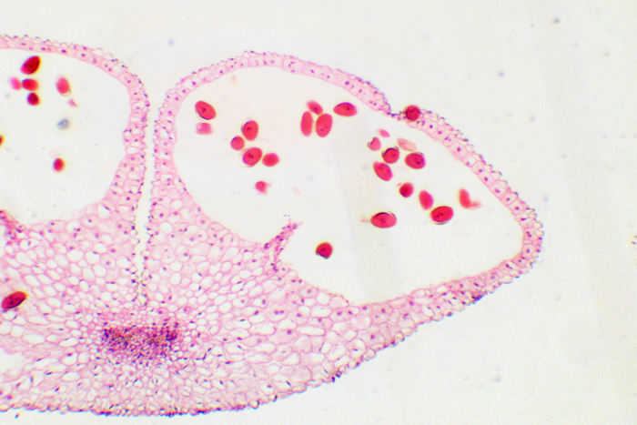
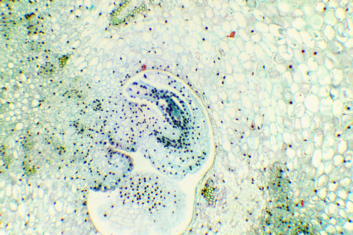
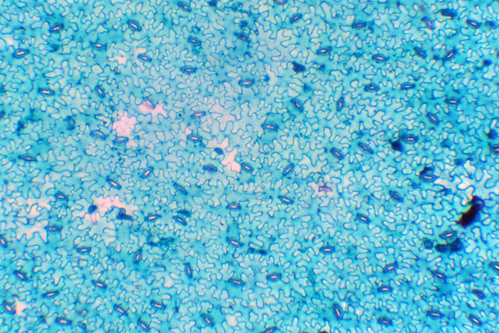
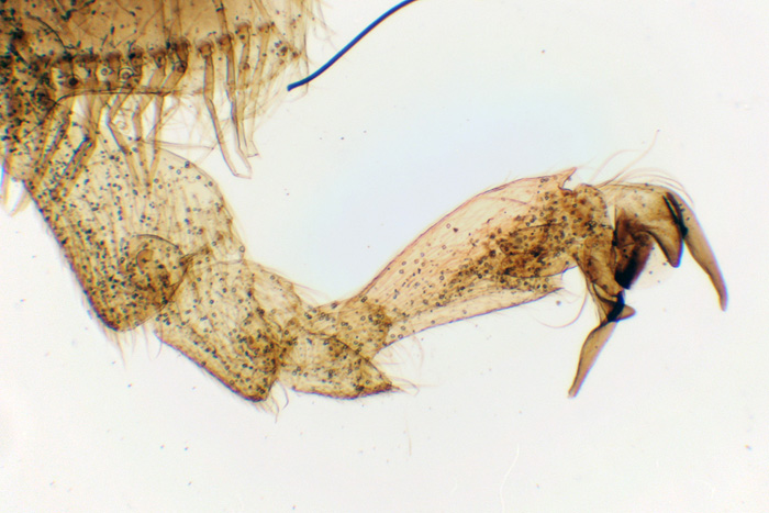
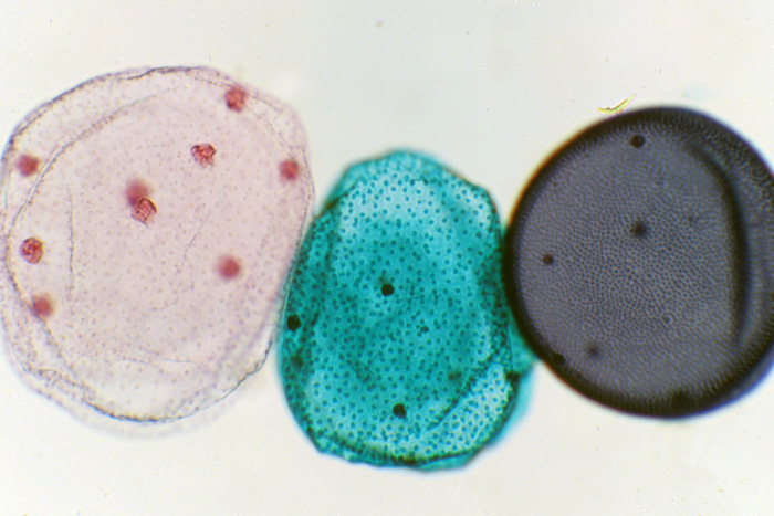
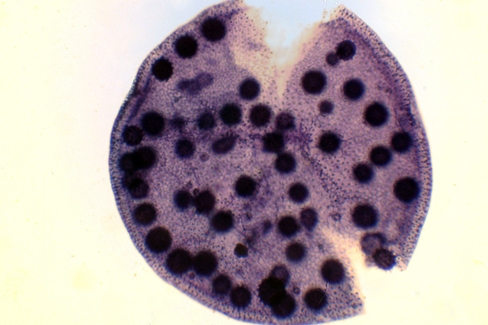
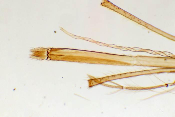
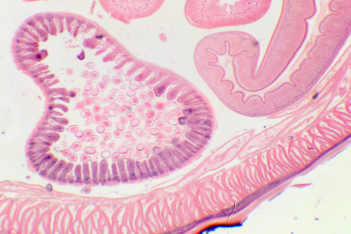
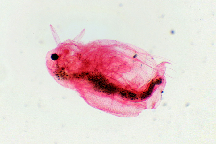
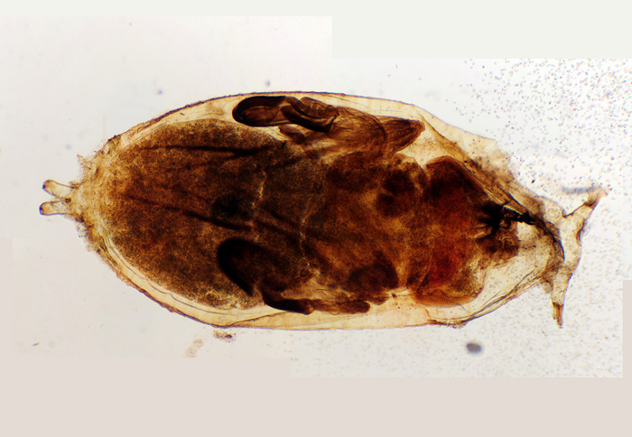
In the kit, you will find the following: onion peel, wheat weevil, root cap, lime twig, anther, plant ovary, camellia, geranium leaf epidermis, bee limb, bee wing, Cyclops, Volvox, Euglena, Paramecium caudatum, earthworm (cross-section), mosquito mouthparts, Ascaris, Daphnia.

The slides are stored in a cardboard box, which protects them from damage during storage and transportation. The legend lists slides from both Levenhuk N18 and Levenhuk N20 Prepared Microscope Slides Set.

The slides are prepared with great care. There are no glue streaks or chips on the slides or cover glass. The specimens are well-placed, without any cracks in the fixing fluid or any significant contamination of the specimens themselves.

The range of slides is rather impressive, so this kit would be a perfect gift for schoolchildren. Below are the images of all the slides in Levenhuk N18 Prepared Microscope Slides Set. The images were made with a Levenhuk 50L NG Microscope and a Canon 350D camera.

levenhuk_experiment_slides_onion_peel.jpg rye_weevil.jpg levenhuk_experiment_slides_root_cap.jpg
Onion peel, 150x Wheat weevil, 60x Root cap, 60x
levenhuk_experiment_slides_linden_twig.jpg levenhuk_experiment_slides_anther.jpg levenhuk_experiment_slides_plant_ovary.jpg
Lime twig, 60x Anther, 60x Plant ovary, 60x
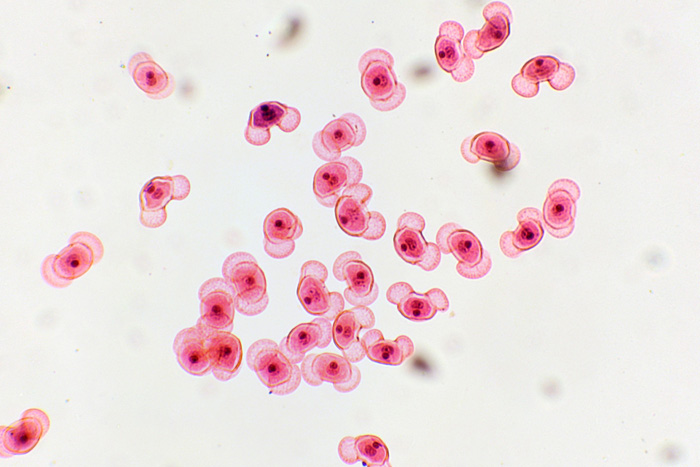
levenhuk_experiment_slides_geranium_leaf_epidermis_60x.jpg  levenhuk_experiment_slides_geranium_leaf_epidermis_150x.jp levenhuk_experiment_slides_pine_pollen.jpg
Geranium leaf
epidermis, 60x
Geranium leaf
epidermis, 150x
Pine pollen, 150x
 levenhuk_experiment_slides_bee_leg.jpg  levenhuk_experiment_slides_cyclops.jpg levenhuk_experiment_slides_cyclops-2.jpg
Bee limb, 60x (it is, of
course, bigger than this;
there's still a lot to study)
Cyclops, 60x Cyclops, 60x
 levenhuk_experiment_slides_bee_wing.jpg  levenhuk_experiment_slides_euglena.jpg levenhuk_experiment_slides_volvox-60x.jpg
Bee wing, 60x Euglena, 600x Volvox, 60x
volvox-150x.jpg  levenhuk_experiment_slides_paramecium_caudatum.jpg  levenhuk_experiment_slides_earthworm.jpg
Volvox, 150x Paramecium caudatum,
150x
Cross-section of
an earthworm, 60x
levenhuk_experiment_slides_mouthparts_mosquito.jpg  levenhuk_experiment_slides_ascaris.jpg levenhuk_experiment_slides_daphnia.jpg
Mouthparts of
a mosquito, 60x
Ascaris, 60x Daphnia, 60x

Now, let’s take a look at  Levenhuk N20 Prepared Microscope Slides Set. It includes slides for biological and physiological studies. The packaging and quality of the slides are on par with Levenhuk N18, which we looked at earlier. However, the “Striated muscles” slide actually has two different specimens in it.

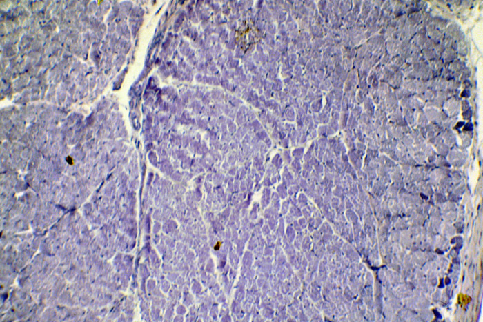
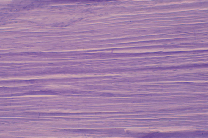
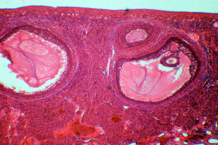
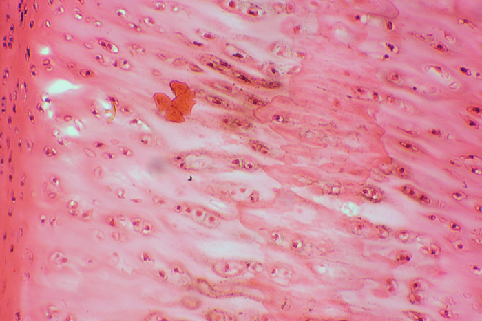
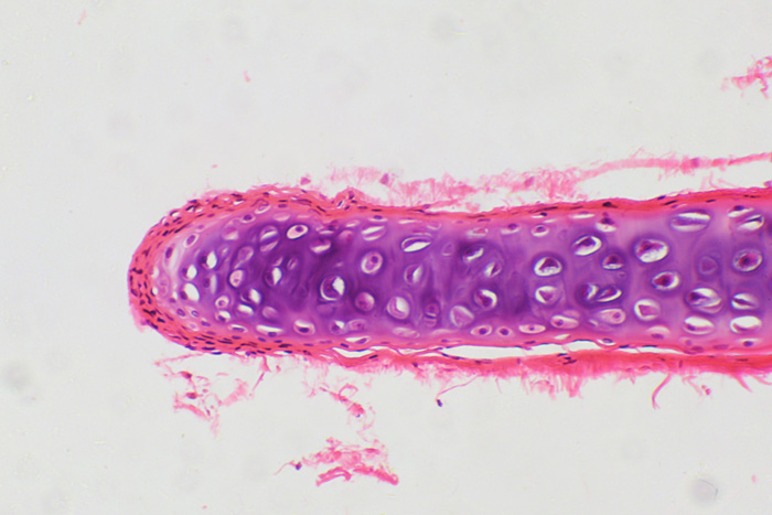
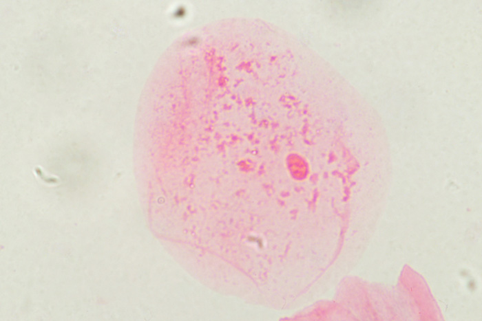
The Levenhuk N20 Prepared Microscope Slides Set includes the following: striated muscles, mammalian sperm cells, cross-section of a nerve, loose connective tissue, mammalian oocyte, nerve cells, hyaline cartilage, smooth muscles, bone tissue, frog blood, human blood, simple epithelium, Drosophila mutation (wingless), Drosophila mutation (black body), normal Drosophila, animal cell, plant cell, mucor mold, fragmentation of the egg, onion root mitosis.

Below are the images of all the slides in  Levenhuk N20 Prepared Microscope Slides Set. The images were made with a LOMO MicMedvo-1 Var. 2-20 microscope and a Canon 350D camera.

 levenhuk_experiment_slides_drosophila_mutation.jpg  levenhuk_experiment_slides_normal_drosophila.jpg  levenhuk_experiment_slides_drosophila_mutation_black_body.jpg
Drosophila mutation
(wingless), 60x
Mosaic image.
Normal Drosophila, 60x
Mosaic image.
Drosophila mutation
(black body), 60x
Mosaic image.
levenhuk_experiment_slides_animal_cell.jpg  levenhuk_experiment_slides_plant_cell.jpg  levenhuk_experiment_slides_mucor_mold_60x.jpg
Animal cell, 150x Plant cell, 150x Mucor mold, 60x
 levenhuk_experiment_slides_mucor_mold_150x.jpg levenhuk_experiment_slides_mucor_mold_600x.jpg  levenhuk_experiment_slides_ovum_cleavage.jpg
Mucor mold
(dark field), 150x
Mucor mold, 600x Fragmentation of the egg, 60x
 levenhuk_experiment_slides_onion_root_mitosis.jpg  levenhuk_experiment_slides_striated_muscles.jpg levenhuk_experiment_slides_striated_muscles2.jpg
Onion root mitosis, 600x Striated muscles, 150x Striated muscles, 150x
 levenhuk_experiment_slides_mammal_spermatozoon.jpg levenhuk_experiment_slides_cross-section_nerve.jpg levenhuk_experiment_slides_fibrous_connective_tissue.jpg
Mammalian sperm cells,
600x
Cross-section
of a nerve, 150x
Loose connective
tissue, 600x
levenhuk_experiment_slides_mammal_ovum.jpg  levenhuk_experiment_slides_nerve_cells.jpg levenhuk_experiment_slides_hyaline_cartilage.jpg
Mammalian oocyte, 60x Nerve cells, 150x Hyaline cartilage, 150x
 levenhuk_experiment_slides_smooth_muscles.jpg  levenhuk_experiment_slides_bone_tissue.jpg  levenhuk_experiment_slides_human_blood.jpg
Smooth muscles, 150x Bone tissue, 150x Human blood, 150x
 levenhuk_experiment_slides_frog_blood_150x.jpg  levenhuk_experiment_slides_frog_blood_600x.jpg  levenhuk_experiment_slides_simple_epithelium.jpg
Frog blood, 150x Frog blood, 600x Simple epithelium, 600x


Any reproduction of the material for public publication in any information medium and in any format is prohibited. You can refer to this article with active link to eu.levenhuk.com.

The manufacturer reserves the right to make changes to the pricing, product range and specifications or discontinue products without prior notice.
View related articles (3)
Levenhuk DTX RC2 Remote Controlled Microscope – Promotional Video
Promotional video of the Levenhuk DTX RC2 Remote Controlled Microscope
Levenhuk DTX 700 Mobi Digital Microscope – Promotional Video
Promotional video of the Levenhuk DTX 700 Mobi Digital Microscope
Levenhuk Rainbow 2L PLUS Moonstone Microscope Promo Video
Promo video of the children's Levenhuk Rainbow 2L PLUS microscope
All articles