
 BG
BG  BY
BY  CY
CY  CZ
CZ  DE
DE  EE
EE  ES
ES  EU
EU  GR
GR  HU
HU  IS
IS  IT
IT  LT
LT  LV
LV  MY
MY  PL
PL  PT
PT  RO
RO  SK
SK  TR
TR  UA
UA MAGUS Bio D240T LCD Biological Digital Microscope
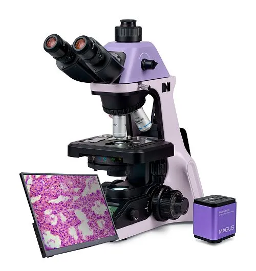
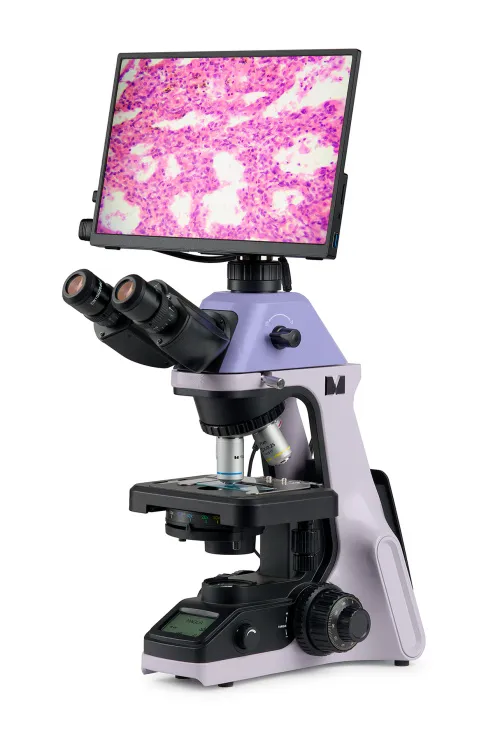
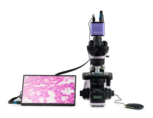
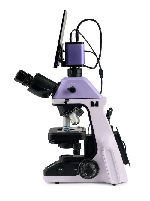


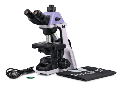
With a camera and a monitor. Magnification: 40–1000x. Trinocular microscope head, coded revolving nosepiece, plan achromatic objectives, 3W LED illuminator, and intelligent lighting control system
| Product ID | 83282 |
| Brand | MAGUS |
| Warranty | 5 years |
| EAN | 5905555019901 |
| Package size (LxWxH) | 17.7x11.8x25.6 in |
| Shipping Weight | 27.7 lb |
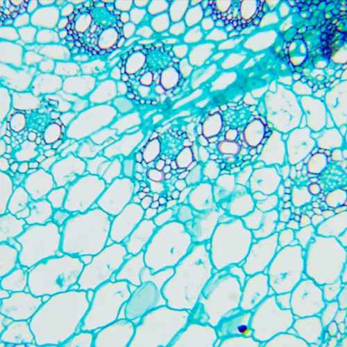
The microscope is suitable for observing transparent and translucent biological samples, such as smears and cross sections using the brightfield microscopy technique in transmitted light. The coded revolving nosepiece maintains a comfortable brightness level when the objectives are changed. The microscope's intelligent lighting control system improves comfort and speed of the researcher’s everyday work. Smart features help students ease into the profession and gain the professional experience they need. Practical aspects play an important role for student microscopes. The size, weight, ease of storage of the cords, and the microscopes themselves are important in everyday use. This microscope is convenient to move around on a table due to its small dimensions and low weight. It does not take up much space in storage. It is equipped with a 2MP camera with an HDMI interface. The digital camera outputs the image directly on the monitor screen with no connection to a computer. The software complements the system with analysis and documentation functions. The monitor in the digital microscope has Full HD resolution.
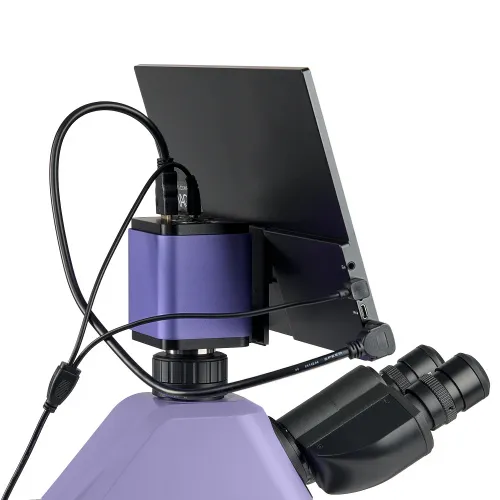
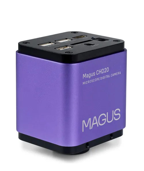
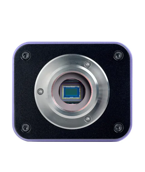
Digital camera
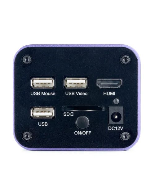
The MAGUS CHD20 Digital Camera is equipped with a 2MP sensor and produces realistic Full HD images at a resolution of 1920x1080px. The camera features high light sensitivity that is suitable for working in fluorescent light. Through the use of RGB primary color mosaic filters, low dark current and a clear image are achieved. The camera uses an HDMI interface to connect directly to a TV, monitor, or projector. In this mode, the camera operates autonomously without a PC. The HDMI interface provides a high and stable transfer rate from the camera to the external screen. An additional USB2.0 interface is provided for connecting the camera to a PC. Video is recorded at 60fps or 50fps depending on the video output interface. The camera combines high FPS and high bandwidth HDMI and, therefore, videos are vivid with no freezes or gaps between frames. At maximum resolution, the image is well-detailed, moving objects are visible without any bugs, and object movement is displayed without delays.
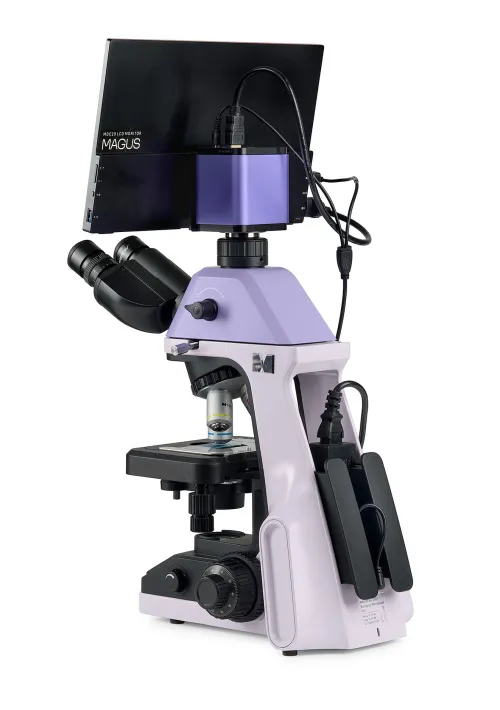
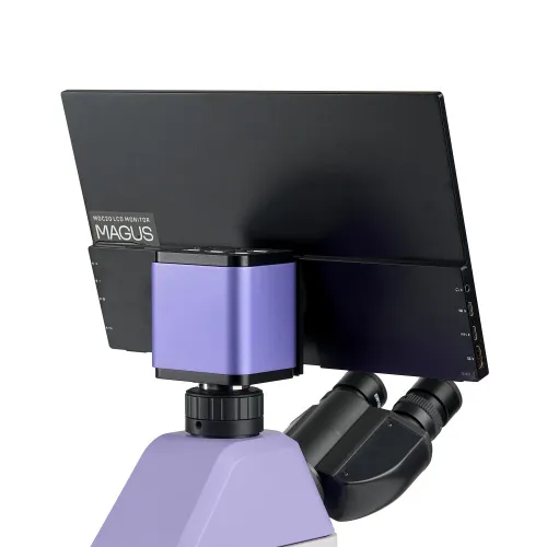
Monitor
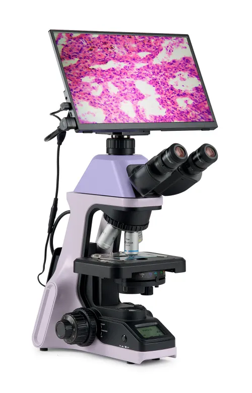
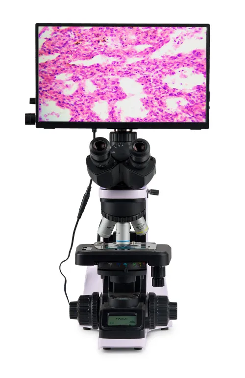
The MAGUS MCD20 Monitor is designed to use a visualization system of the MAGUS microscope. It is connected to a camera mounted on the microscope to display real-time images. It is compatible with MAGUS HDMI cameras operating in Full HD resolution. The screen diagonal is 13.3 inches. The IPS sensor delivers a bright image with large viewing angles: If you look at the monitor at an angle, there is no color distortion. The display can be placed on a folding stand on a table or mounted directly on the camera, whichever is more convenient for the user.
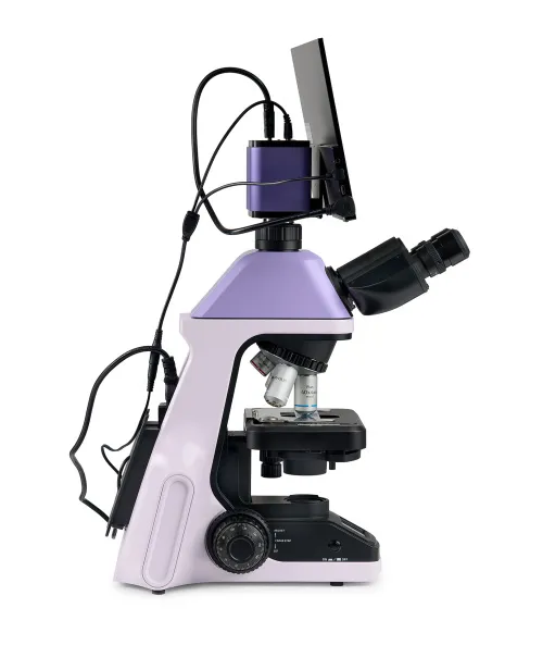
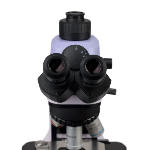
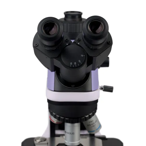
Microscope head
Trinocular head with infinity-corrected optics. The eyepiece tubes are 360° rotatable. The user can adjust the eyepiece height to suit their individual stature. The digital camera is mounted in the trinocular tube. Beam splitting ratio: 0/100 and 100/0. The kit includes 10x/20mm eyepieces with long eye relief. Flat rubber eyecups without protruding parts protect the optics of glasses from scratches. Diopter adjustment is done directly on the microscope: Diopter adjustment rings are located on both eyepiece tubes.
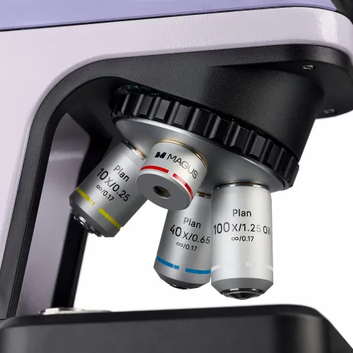
Revolving nosepiece
The coded revolver for four objectives is oriented toward the interior: The user can see the objective inserted into the optical path, and the space above the stage is free.
Maintaining comfortable brightness levels when switching magnifications
The objectives of different magnifications transmit light with different levels of intensity, and so each time you change objectives, the brightness of the light must be adjusted. Switching from a higher to a lower magnification objective causes eye fatigue, as the image brightness in the eyepieces increases sharply. The MAGUS Bio D240T LCD, which is equipped with intelligent brightness control, solves this problem. The microscope remembers the brightness for each objective that the user has selected and automatically sets this brightness when turning the nosepiece. Intelligent control reduces the time required to adjust brightness. MAGUS Bio D240T LCD increases user comfort and saves time even when the given work requires frequent magnification changes.
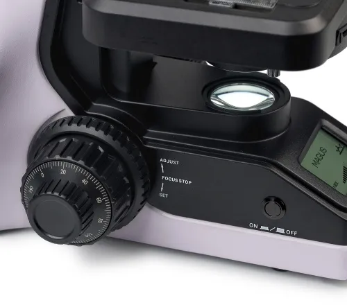
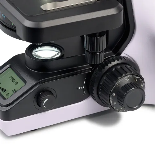
Focusing mechanism
Coaxial coarse and fine focusing knobs are located at the bottom of the microscope body on both sides of the stand. The user can place their hands on the table and take a relaxed pose while observing. The focus adjustment is smooth and effortless. The coarse focus lock knob helps you quickly adjust the microscope after changing the object of study. The knob is located on the left side of the microscope on the same axis as the focusing mechanism. The ring on the right side adjusts the tension of the coarse focusing travel. The user adjusts the comfortable tension for work.
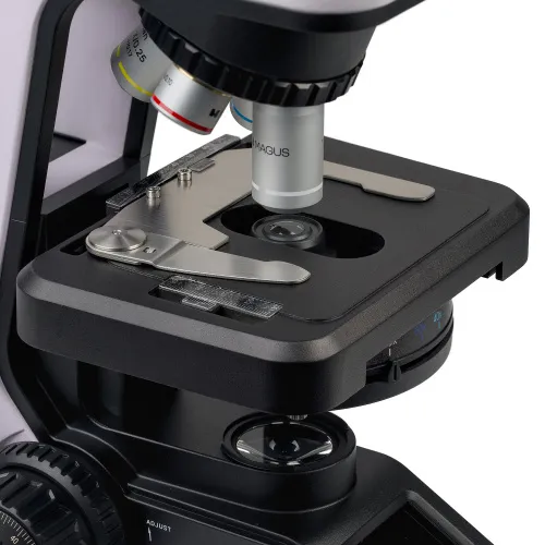
Stage
The stage has no X-axis rack and pinion, which improves ergonomics. The belt-driven mechanism allows for smooth movement of the specimen. The specimen holder is secured with two screws and can be easily removed during manual scanning.
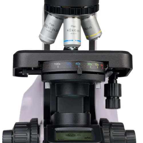
Abbe condenser
An Abbe immersion condenser with N.A. 1.25 is fixed with two screws under the stage. The height of the condenser position is set at the factory and does not require adjustment by the user. The condenser is centered relative to the optical axis. Locking the condenser position eliminates the risk of accidentally changing the correct setup. The pre-configured condenser makes it easier for students to operate the microscope and it frees up more time for science. The condenser knob adjusts the iris aperture diaphragm. The color marking corresponds to the objective magnification. To achieve a contrast image on each objective, it is recommended to set the aperture adjustment knob to a position that corresponds to the number designation of the objective.
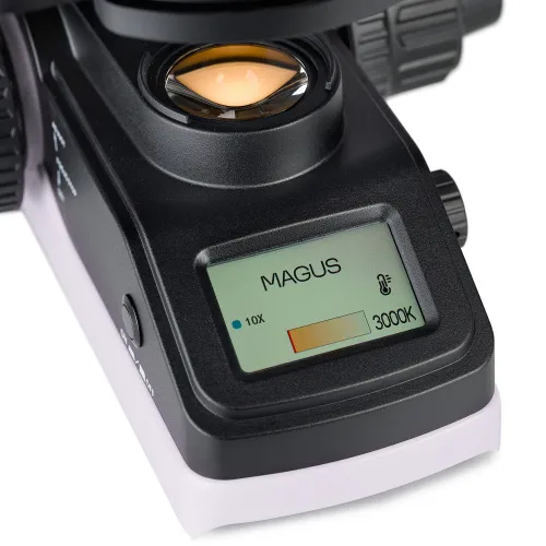
Light source
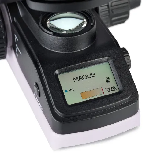
The transmitted light illuminator contains a 3W LED. The microscope illuminator has the function of adjusting the color temperature in the range of 3000K to 7000K. The user selects a color temperature that is comfortable for their eyes, and they can easily change it with just one button when, for example, examining an object requires changing the light. The LED lifetime is 50,000 hours.
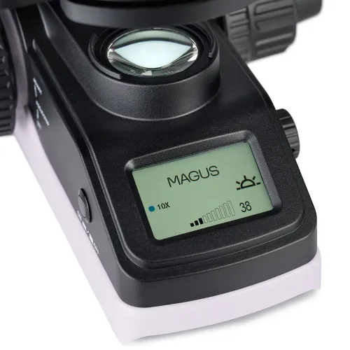
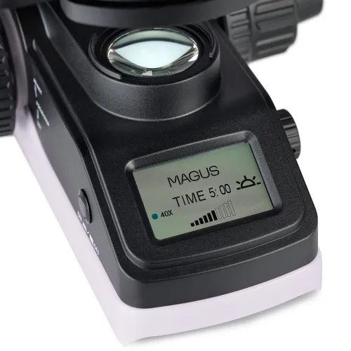
Status LCD screen
The LCD screen on the base of the microscope displays the objective magnification, brightness and color temperature of the light source, and operating mode (“sleep” and “eco”). Using the screen and two knobs, the microscope user adjusts the brightness, selects the color temperature, locks the brightness adjustment, and sets the sleep mode and auto-off timer.
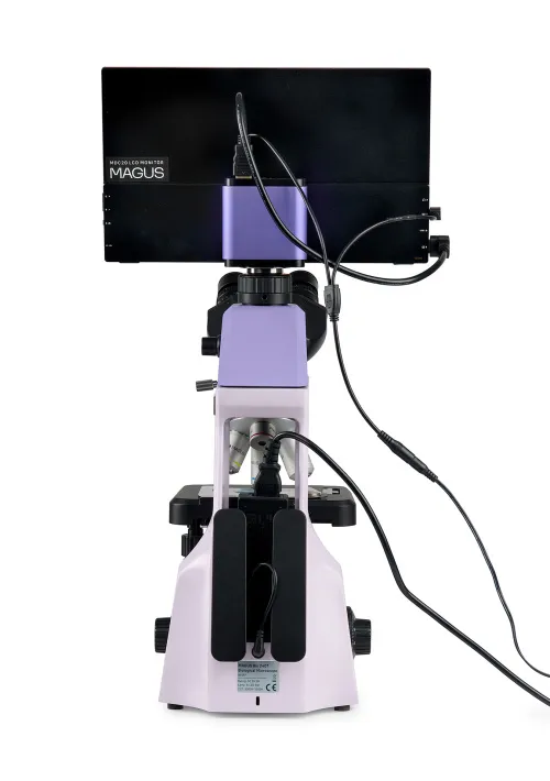
Ergonomic design
The sides of the “window” in the stand form handles for carrying the microscope with two hands. The design for the hidden placement of the power adapter and power cord improves the workplace esthetic and safety of carrying the microscope as well as simplifies storage of the device.
Alternative models
The MAGUS Bio D240T is an alternative for the following models: Nikon ECLIPSE Ei, Olympus CX 23.
Microscope features:
- Observations of transparent and translucent samples in brightfield in transmitted light
- Trinocular head with a vertical tube for installing a digital camera, height adjustment to adjust to the observer, and light beam splitting 0/100 and 100/0
- Coded revolving nosepiece: The brightness of the light source is set automatically depending on the selected objective
- Pre-configured condenser with color-coded objective magnification allows for quick and precise aperture setting
- Transmitted light illuminator: energy-saving 3W LED with a lifetime of up to 50.000 hours
- Intelligent lighting control system: automatic brightness selection when changing objectives and color temperature adjustment, brightness adjustment lock, auto-off timer, and LCD status screen
- Stage without a positioning rack for convenient operation
- Ergonomic stand with carrying handles and the hidden placement of the power cord and power adapter
- Compact and lightweight design makes it easy to store the microscope on high shelves
Camera features:
- The camera operates autonomously without a connection to a PC via HDMI interface. Can be connected to a PC via USB2.0 interface
- The camera resolution of 1920x1080px is the best choice for displaying images on a Full HD monitor or TV
- 60fps or 50fps, depending on the video output interface for observing moving objects, recording video, and moving the preparation without jerkiness or lags
- SONY Starvis Exmor color CMOS backlit sensor provides low noise level and high light sensitivity even in low-light conditions. You will get clearer, brighter, and more color-saturated images
- Software with photo, video recording, editing, external display functions, linear and angular measurements
Monitors features:
- Full HD resolution is the most popular and convenient resolution for working with digital images
- The IPS sensor produces a bright and saturated picture with wide vertical and horizontal viewing angles
- The monitor can be mounted on the camera or table to suit the user’s preferences
Package:
- MAGUS CHD20 digital camera (digital camera, HDMI cable (1.5m), USB 2.0 cable (2m), USB mouse, 32GB SD memory card, 12V/1A (Euro) power adapter, USB flash drive with drivers and software, user manual, and warranty card)
- MAGUS MCD20 Monitor
- Stand with the transmitted light source, focusing mechanism, stage, condenser, and revolving nosepiece
- Abbe condenser
- Trinocular microscope head
- Plan achromatic objective Plan 4x/0.10 ∞/0.17
- Plan achromatic objective Plan 10x/0.25 ∞/0.17
- Plan achromatic objective Plan 40x/0.65 (spring-loaded)
- Plan achromatic objective Plan 100x/1.25 (oil) ∞/0.17 (spring-loaded)
- 10x/20mm eyepiece with long eye relief (2 pcs.)
- Eyecup (2 pcs.)
- 0.55x C-mount adapter
- Color filter
- Microscope power adapter and power cord
- Dust cover
- User manual and warranty card
Available on request:
- 10x/20mm eyepiece with a scale (D 23.2mm)
- 10x/20mm eyepiece with a center field pointer (D 23.2mm)
- Plan achromatic objective Plan 20х/0.40 ∞/0.17
- Plan achromatic objective Plan 60х/0.80 ∞/0.17
- 1х C-mount adapter
- Calibration slide
- Set of color filters (blue, green, yellow, frosted glass)
| Product ID | 83282 |
| Brand | MAGUS |
| Warranty | 5 years |
| EAN | 5905555019901 |
| Package size (LxWxH) | 17.7x11.8x25.6 in |
| Shipping Weight | 27.7 lb |
| Type | biological, light/optical |
| Microscope head type | trinocular |
| Head | Gemel head (Siedentopf, 360° rotation), beam splitting 0/100, 100/0 |
| Head inclination angle | 30 ° |
| Magnification, x | 40 — 1000 |
| Magnification, x | 40–1000 basic configuration |
| C-mount adapter magnification, x | 0.55 |
| Eyepiece tube diameter, in | 0.9 |
| Eyepiece tube diameter, mm | 23.2 mm (third vertical tube) |
| Eyepieces | 10x/20mm, long eye relief (*optional: 10x/20 with a scale, 10x/20 with a pointer) |
| Objectives | infinity plan achromatic: 4x/0.10; 10x/0.25; 40xs/0.65; 100xs/1.25 oil; parfocal distance: 45mm |
| Revolving nosepiece | 4 objectives, coded |
| Interpupillary distance, in | 1.9 — 3 |
| Interpupillary distance, mm | adjustable |
| Stage, mm | 180x130 |
| Stage moving range, mm | 74/30 |
| Stage features | two-axis mechanical stage, without a positioning rack |
| Eyepiece diopter adjustment, diopters | ±5D on both tubes |
| Eyepiece diopter adjustment | ✓ |
| Condenser | Abbe condenser N.A. 1.25 with an adjustable aperture diaphragm and color-coded objective magnification |
| Diaphragm | adjustable aperture |
| Focus | coaxial, coarse (17mm, 37.7mm/circle, with a lock knob and tension adjusting knob) and fine (0.002mm, 0.2mm/circle) |
| Illumination | LED |
| Power supply | 100–240V, 50/60Hz (US type plug adapter is not included), AC network, the AC/DC power adapter is located in a special socket on the back of the stand |
| Light source type | 3W LED, with color temperature adjustment (3000–7000K) |
| Light filters | yes |
| Additional | automatic brightness adjustment when switching objectives, eco mode, sleep mode, status display on LCD screen |
| User level | experienced users, professionals |
| Assembly and installation difficulty level | complicated |
| Application | laboratory/medical |
| Illumination location | lower |
| Research method | bright field |
| Pouch/case/bag in set | dust cover |
| Sensor | Sony Exmor/Starvis CMOS |
| Color/monochrome | color |
| Megapixels | 2 |
| Maximum resolution, pix | 1920x1080 |
| Pixel size, μm | 3.75x3.75 |
| Interface connectors | HDMI 1.4, USB 2.0 |
| Memory card | SD up to 32GB |
| Ability to connect additional equipment | USB mouse, flash stick (USB) |
| Light sensitivity | 1175mV with 1/30s |
| Signal/noise ratio | 0.15mV at 1/30s |
| Exposure time | 0.04ms–1000ms |
| Video recording | ✓ |
| Video recording | yes |
| Frame rate, fps at resolution | 50@1920x1080 (USB), 60@1920x1080 (HDMI) |
| Place of installation | trinocular tube, eyepiece tube instead of an eyepiece |
| Image format | *.jpg, *.tif |
| Video format | *.h264/*.h265, *.mp4 |
| Spectral range, nm | 380–650 (IR-filtered) |
| Shutter type | ERS (electronic rolling shutter) |
| Software | HDMI: built-in; USB: MAGUS View |
| System requirements | Windows 8/10/11 (32bit and 64bit), Mac OS X, Linux, up to 2.8GHz Intel Core 2 or higher, minimum 4GB RAM, USB2.0 port, RJ45, 19" or larger display |
| Mount type | C-mount |
| Body | aluminum |
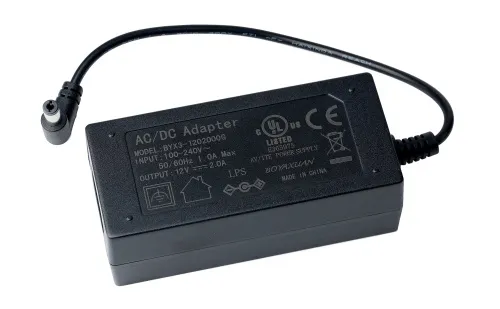
| Camera power supply | AC adapter 12V, 1A (US type plug adapter is not included) |
| Specifications of the AC power adapter | input: AC voltage 100–240V, 50/60Hz, output: DC voltage 12V/1A |
| Camera operating temperature range, °F | -10...+50 |
| Operating humidity range, % | 30 — 80 |
| Type of matrix | IPS |
| Display diagonal, inch | 13.3 |
| Display resolution, px | 1920x1080 (Full HD) |
| Aspect ratio | 16:9 |
| Brightness, cd/m² | 400 |
| Number of displayed colors | 16.7 m |
| Contrast | 1000:1 |
| Horizontal/vertical viewing angle, ° | 178/178 |
| Size of the visible screen area (WxH), mm | 295x165 |
| Pixel pitch (WxH), mm | 0.154x0.154 |
| Frequency of optical source, Hz | 60 |
| Type of matrix backlight | LED |
| LED backlight lifetime, h | 50000 |
| Operating temperature range, °F | 5...+131 |
| Operating humidity range, % | 10 — 90 |
| Power supply | AC 110–220V (US type plug adapter is not included), DC 5–12V/1A (Type-C) |
| Power consumption, W | 12 (max.) |
We have gathered answers to the most frequently asked questions to help you sort things out
Find out why studying eyes under a microscope is entertaining; how insects’ and arachnids’ eyes differ and what the best way is to observe such an interesting specimen
Read this review to learn how to observe human hair, what different hair looks like under a microscope and what magnification is required for observations
Learn what a numerical aperture is and how to choose a suitable objective lens for your microscope here
Learn what a spider looks like under microscope, when the best time is to take photos of it, how to study it properly at magnification and more interesting facts about observing insects and arachnids
This review for beginner explorers of the micro world introduces you to the optical, illuminating and mechanical parts of a microscope and their functions
Short article about Paramecium caudatum - a microorganism that is interesting to observe through any microscope
