
 BG
BG  BY
BY  CY
CY  CZ
CZ  DE
DE  EE
EE  ES
ES  EU
EU  GR
GR  HU
HU  IS
IS  IT
IT  LT
LT  LV
LV  MY
MY  PL
PL  PT
PT  RO
RO  SK
SK  TR
TR  UA
UA MAGUS Bio DH240 Biological Digital Microscope
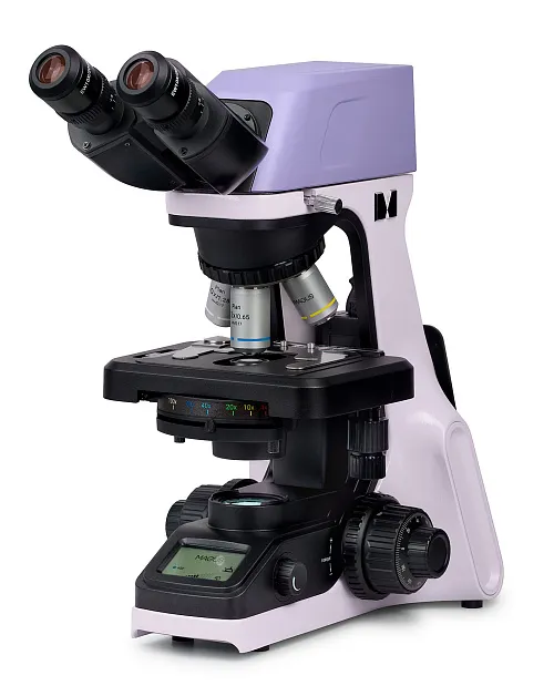


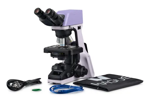
Magnification: 40–1000x. Binocular head with a built-in 8MP digital camera, coded revolving nosepiece, plan achromatic objectives, 3W LED illuminator, intelligent lighting control system
| Product ID | 83478 |
| Brand | MAGUS |
| Warranty | 5 |
| EAN | 5905555019444 |
| Package size (LxWxH) | 17.7x11.8x25.2 in |
| Shipping Weight | 21.8 lb |
The MAGUS Bio DH240 microscope is designed to work with transparent and translucent biological samples in the brightfield method. The device is equipped with a binocular head with a built-in digital camera. The 4-objective revolving nosepiece is equipped with an intelligent lighting control system. The selected operating settings are displayed on the built-in LCD screen.
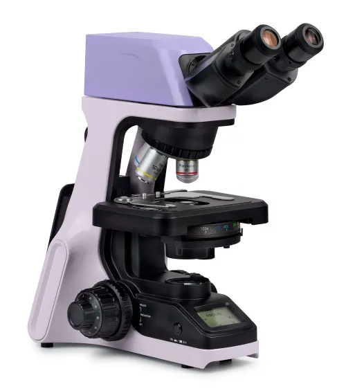
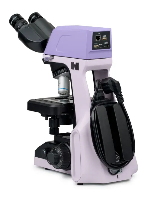
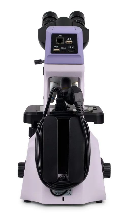
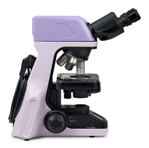
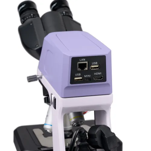
The MAGUS Bio DH240 model is fairly compact. It is lightweight and small in size, and the ergonomic design of the body makes it easy to store and transport. All of the cords and the power adapter are hidden on the back of the microscope, and you can use the side openings of the stand as handles for carrying it.
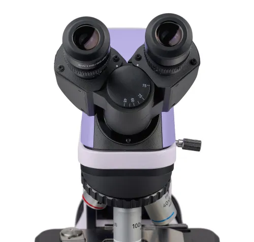
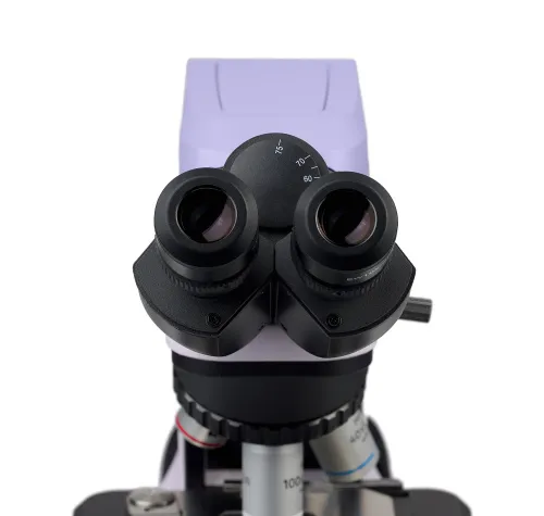
The binocular head also adds to the design: The digital camera is built into it and does not take up extra space. Both tubes rotate 360° around their axis, and so you can adjust the eye relief according to your requirements. The microscope is equipped with 10x/20mm eyepieces, and includes flat rubber eyecups, which will be handy for users with glasses to prevent the lenses touching the eyepieces and to avoid damage. The optics are infinity-corrected. There are diopter adjustment rings on the tubes of the microscope that adjust to the user’s visual acuity.
A digital camera built on an 8MP matrix produces images with a resolution of 4K (3840x2160px). It perfectly conveys all of the nuances of the object of observation. The optimal combination is obtained with 4x and 10x objectives. The camera can take photographs or videos of static and inactive specimens. It can be used to adjust the focus of the microscope. You can transfer images to your computer via Wi-Fi.
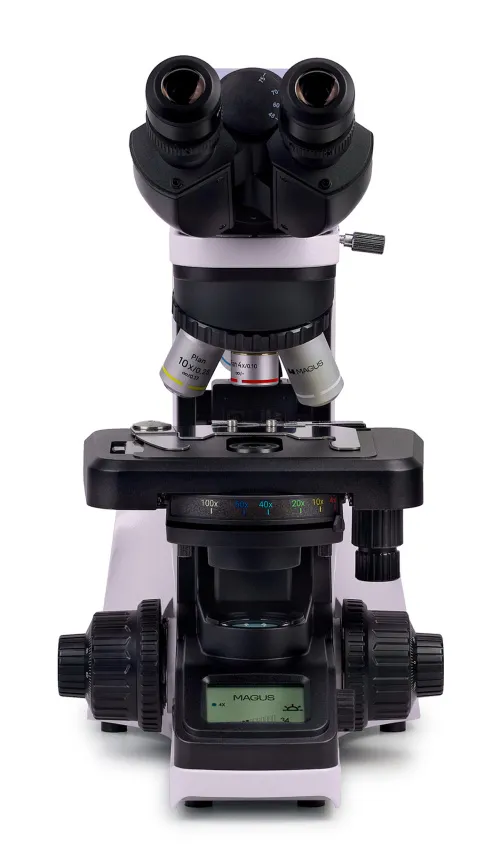
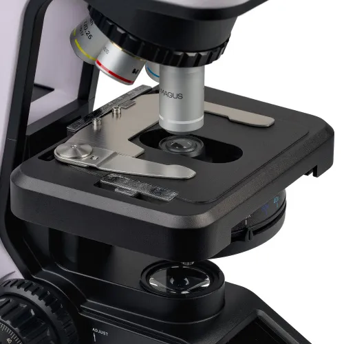
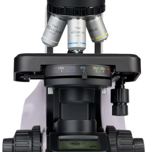
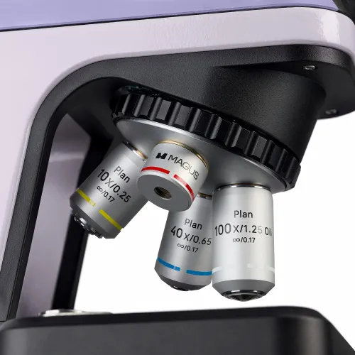
The revolving nosepiece of the MAGUS Bio DH240 model is designed for 4 objectives. It is oriented “toward the interior”: The working objective is located directly in front of the user, while the rest are pointed in the opposite direction. This design frees up space in front of the user, making work more convenient. Coded revolving nosepiece: When you switch the objective, the illumination of the specimen changes automatically. Since objectives with different magnifications have different light transmission, switching objectives means the intensity of the illumination in the eyepiece greatly fluctuates. This strains the eyes, and the researcher has to spend time adjusting the brightness of the light each time. The intelligent lighting system remembers the user’s settings for each objective, and when you turn the revolver, the light automatically adjusts to the right intensity. This is especially convenient if you frequently change the microscope magnification during your work.
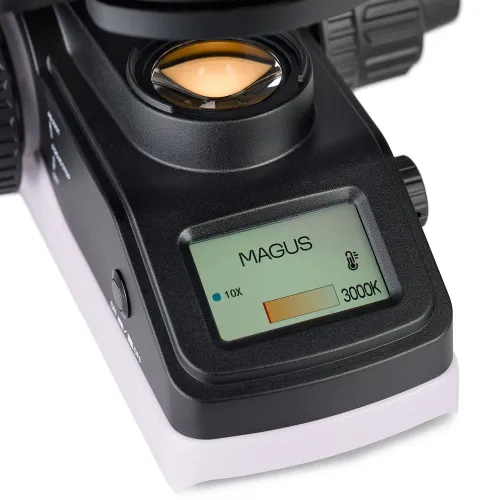
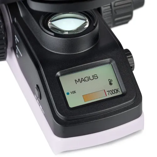
The microscope uses a 3W LED as the light source. The illuminator creates transmitted light with the ability to change color temperature: The user can set the desired value in the range of 3000–7000K. The LED is designed to last for 50,000 hours.
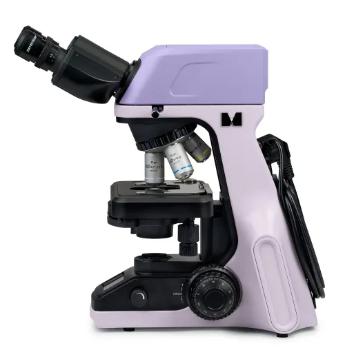
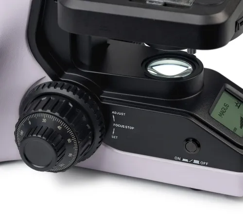
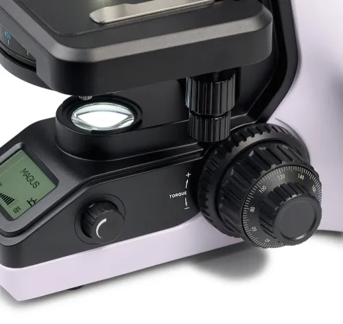
Two focusing knobs are located on the same axis on opposite sides of the microscope and are used for coarse and fine adjustment. In addition, the microscope is equipped with a coarse focus locking handle that is used after changing the objective, and a ring for adjusting the tension of the coarse focus travel. All of the adjustment mechanisms are located in the lower part of the microscope body. To operate it, the user can rest their hands on the table and take a comfortable position, which is especially important during prolonged study.
The specimen stage is especially ergonomic. It is equipped with a belt drive mechanism, and so the specimen moves smoothly along the surface of the stage. The specimen holder is secured with 2 screws and can be easily removed. There is no positioning rack.
The Abbe condenser in this model also comes with some features. It is fixed at the required height and located in the center of the optical axis of the microscope: It cannot be accidentally moved, which is especially important for the work of novice researchers in a common space. A knob is used to adjust the iris diaphragm, which is set according to the markings on the condenser. Each marking corresponds to a specific objective magnification, and the knob must be moved to the position corresponding to the working objective.
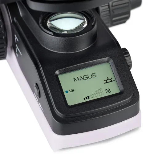
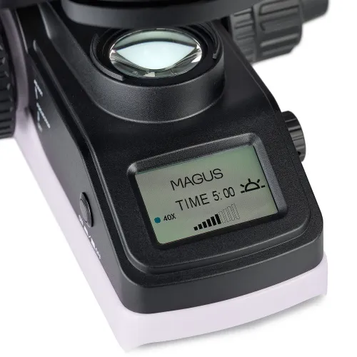
You can track all of the settings on the built-in LCD display, which is another feature of this MAGUS microscope model. The screen displays the selected brightness of the illuminator and its color temperature, the magnification of the working objective, and the operating mode – most of these functions are configured using the screen.
Key features:
- Works with transparent and translucent specimens in the brightfield method
- The binocular head has a built-in digital camera
- Both tubes rotate 360°, and there is a diopter adjustment system
- Revolving nosepiece for 4 objectives with intelligent lighting control system
- Selected operating parameters are displayed on the LCD screen
- Compact and lightweight device with an ergonomic body: The cords and adapter are hidden, and the body design is convenient for storage and transportation
- Specimen stage with a belt drive mechanism that does not have a positioning rack
- The condenser does not require additional adjustment; the aperture diaphragm is adjusted according to the magnification markings of the objectives
The kit includes:
- Stand with transmitted light source, focusing mechanism, stage, condenser mount, and revolving nosepiece
- Abbe condenser
- Binocular head with a built-in digital camera
- Plan achromatic objective Plan 4x/0.10 ∞/0.17
- Plan achromatic objective Plan 10x/0.25 ∞/0.17
- Plan achromatic objective Plan 40x/0.65 ∞/0.17 (spring-loaded)
- Plan achromatic objective Plan 100x/1.25 (oil) ∞/0.17 (spring-loaded)
- Eyepiece 10x/20mm with long eye relief (2 pcs.)
- Eyepiece eyecup (2 pcs)
- Light filter
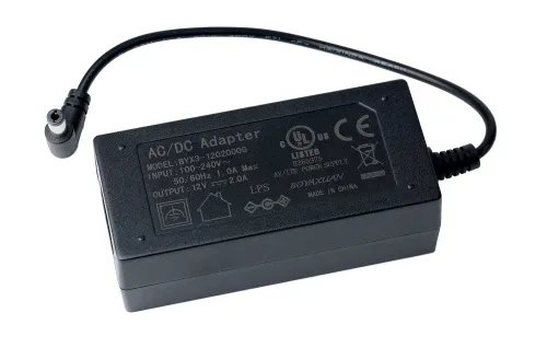
- Microscope power adapter and power cord
- Dust cover
- User manual and warranty card
Available on request:
- 10x/20mm eyepiece with a scale (D 23.2mm)
- 10x/20mm eyepiece with a center field pointer (D 23.2mm)
- Plan achromatic objective Plan 20х/0.40 ∞/0.17
- Plan achromatic objective Plan 60х/0.80 ∞/0.17
- 0.37x C-mount eyepiece adapter
- 0.5x C-mount eyepiece adapter
- 0.75x C-mount eyepiece adapter
- 1х C-mount eyepiece adapter
- Digital camera
- Calibration slide
- Monitor
- Set of color filters (blue, green, yellow, frosted glass)
| Product ID | 83478 |
| Brand | MAGUS |
| Warranty | 5 |
| EAN | 5905555019444 |
| Package size (LxWxH) | 17.7x11.8x25.2 in |
| Shipping Weight | 21.8 lb |
| Microscope specifications | |
| Type | biological, light/optical, digital |
| Microscope head type | binocular |
| Head | Gemel head (Siedentopf, 360° rotation) |
| Head inclination angle | 30 ° |
| Magnification, x | 40 — 1000 |
| Eyepiece tube diameter, in | 0.9 |
| Eyepieces | 10x/20mm, long eye relief (*optional: 10x/20 with a scale, 10x/20 with a pointer) |
| Objectives | plan achromatic, infinity corrected: 4x/0.10; 10x/0.25; 40x/0.65; 100x/1.25 (oil); parfocal height: 45mm (*optional: 20х/0.40; 60x/0.80) |
| Revolving nosepiece | 4 objectives, coded |
| Interpupillary distance, in | 1.9 — 3 |
| Stage, mm | 180x130 |
| Stage moving range, mm | 74/30 |
| Stage features | two-axis mechanical stage, without a positioning rack |
| Eyepiece diopter adjustment, diopters | ±5D on both tubes |
| Eyepiece diopter adjustment | ✓ |
| Condenser | Abbe condenser N.A. 1.25 with an adjustable aperture diaphragm and color-coded objective magnification |
| Diaphragm | adjustable aperture |
| Focus | coaxial, coarse (17mm, 37.7mm/circle, with a lock knob and tension adjusting knob) and fine (0.002mm, 0.2mm/circle) |
| Illumination | LED |
| Brightness adjustment | ✓ |
| Power supply | 100–240V, 50/60Hz (US type plug adapter is not included), AC network, the AC/DC power adapter is located in a special socket on the back of the stand |
| Light source type | 3W LED, with color temperature adjustment (3000–7000K) |
| Light filters | yes |
| Additional | automatic brightness adjustment when switching objectives, eco mode, sleep mode, status display on LCD screen |
| Ability to connect additional equipment | darkfield slider, phase contrast device (condenser and objectives)phase contrast device (slider and objectives), polarization devices (polarizer and analyzer) |
| User level | experienced users, professionals |
| Application | laboratory/medical |
| Illumination location | lower |
| Research method | bright field |
| Digital camera included | ✓ |
| Pouch/case/bag in set | dust cover |
| Camera specifications | |
| Color/monochrome | color |
| Megapixels | 8 |
| Maximum resolution, pix | 3840x2160 |
| Sensor size | 1/2.5" (6.22x3.50mm) |
| Pixel size, μm | 1,62x1,62 |
| Interface connectors | Wi-Fi |
| Light sensitivity | 236mV at 1/30s/0.1mV at 1/30s |
| Exposure time | 0.244ms–15s |
| Video recording | ✓ |
| Video recording | yes |
| Frame rate, fps at resolution | 16@1920x1080, 4@3840x2160 |
| Usage location | built-in |
| Camera power supply | main cord |
and downloads
We have gathered answers to the most frequently asked questions to help you sort things out
Find out why studying eyes under a microscope is entertaining; how insects’ and arachnids’ eyes differ and what the best way is to observe such an interesting specimen
Read this review to learn how to observe human hair, what different hair looks like under a microscope and what magnification is required for observations
Learn what a numerical aperture is and how to choose a suitable objective lens for your microscope here
Learn what a spider looks like under microscope, when the best time is to take photos of it, how to study it properly at magnification and more interesting facts about observing insects and arachnids
This review for beginner explorers of the micro world introduces you to the optical, illuminating and mechanical parts of a microscope and their functions
Short article about Paramecium caudatum - a microorganism that is interesting to observe through any microscope
