BG
BG  BY
BY  CY
CY  CZ
CZ  DE
DE  EE
EE  ES
ES  EU
EU  GR
GR  HU
HU  IS
IS  IT
IT  LT
LT  LV
LV  MY
MY  PL
PL  PT
PT  RO
RO  SK
SK  TR
TR  UA
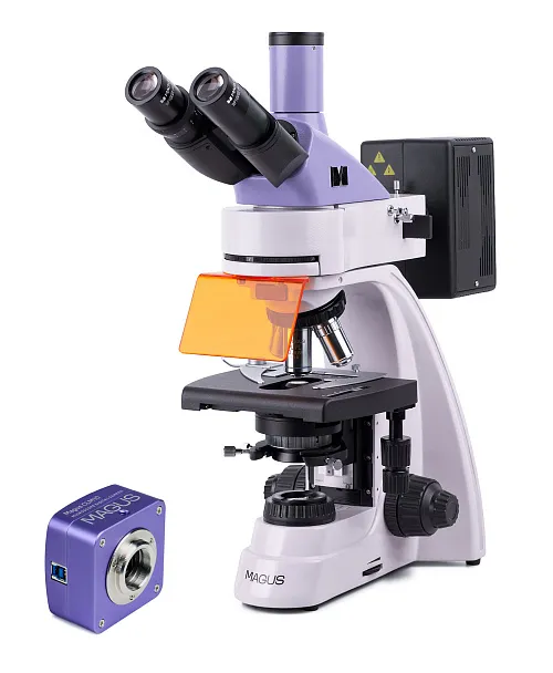
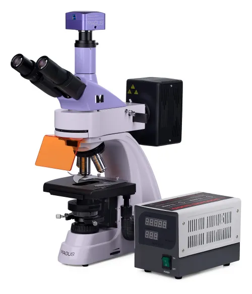
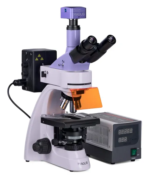
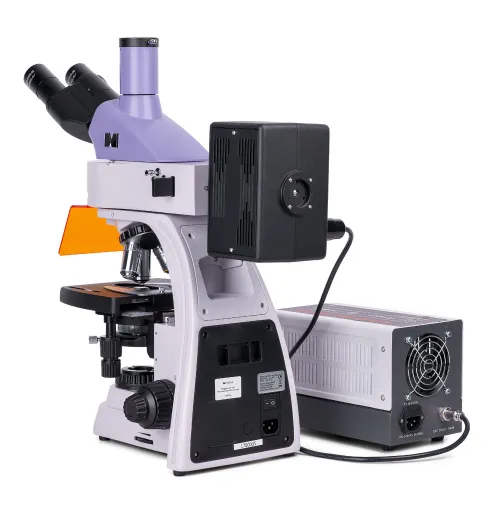
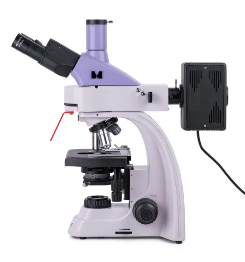
UA MAGUS Lum D400 Fluorescence Digital Microscope
With a camera. Magnification: 40–1000x.Trinocular head, plan achromatic, and plan fluorite objectives, reflected light source – 100W mercury lamp, transmitted light source – 30W halogen lamp, Köhler illumination in reflected and transmitted light
| Product ID | 83016 |
| Brand | MAGUS |
| Warranty | 5 |
| EAN | 5905555018157 |
| Package size (LxWxH) | 17.7x11.8x37.8 in |
| Shipping Weight | 37.7 lb |
Fluorescence microscope MAGUS Lum 400 is designed for diagnostics and research by methods of fluorescence (reflected light) and brightfield (transmitted light) microscopy. Darkfield, phase contrast, and polarization techniques can be used with optional equipment. Areas of application of fluorescence microscope: DNA analysis, study of pathogens, sanitary and epidemiological surveillance, forensic examination, etc.
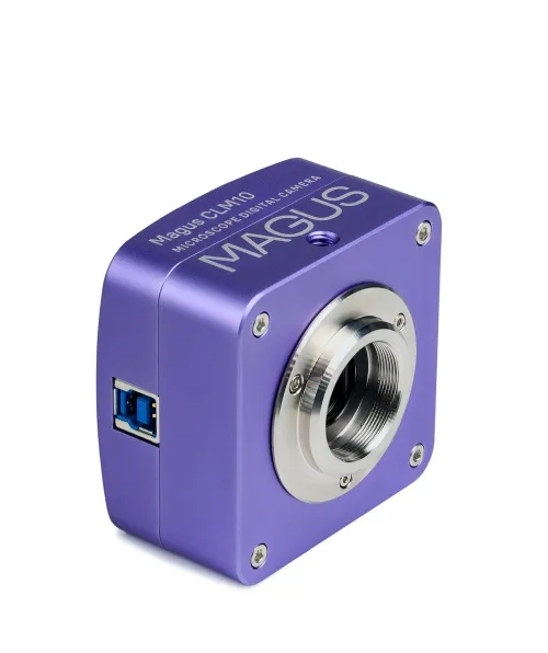
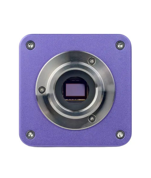
Digital camera
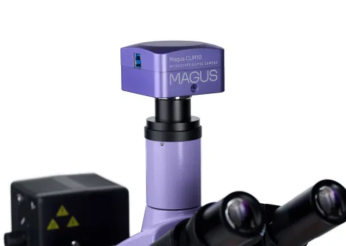
The MAGUS CLM10 digital camera is designed for use in fluorescence and darkfield microscopy techniques. The camera is equipped with a 2.3MP sensor and produces realistic images at a resolution of 1920х1200px. It is recommended for 40x, 60x, and 100x objectives. When observing with medium magnification objectives, the camera will show finer details. Video is recorded at 120fps at maximum resolution. Videos are smooth, with soft and subtle transitions between frames. The movement of the object is displayed in real time with no delays. The camera makes it easy to work with objects that are moving and is suitable for classroom demonstrations. The camera is equipped with a USB3.0 interface. The data transfer speed is 10 times faster than USB2.0 cameras. The high-speed camera is recommended for professional laboratories, research, or university training.
Optics
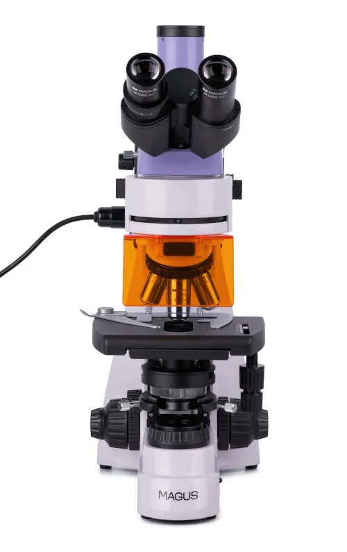
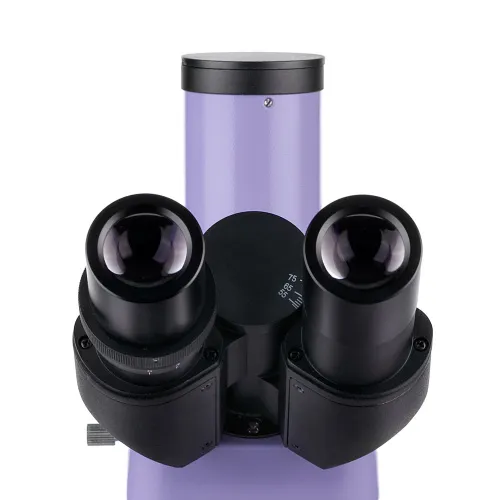
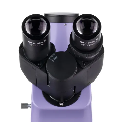
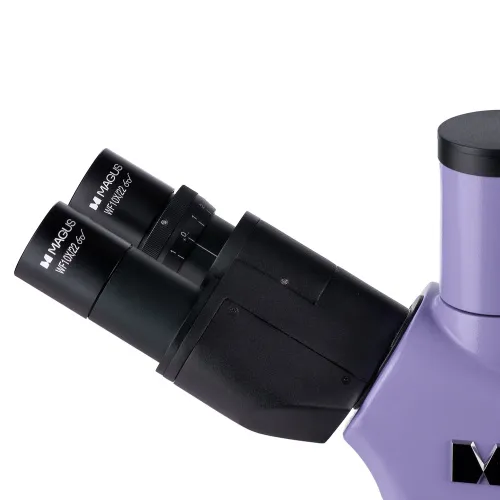
This is a trinocular microscope: The binocular head is 30° inclined and the digital camera (not included) is installed in the trinocular tube. As the eyepiece tubes are 360° rotatable, it is easy to adjust them to fit the height of the user.
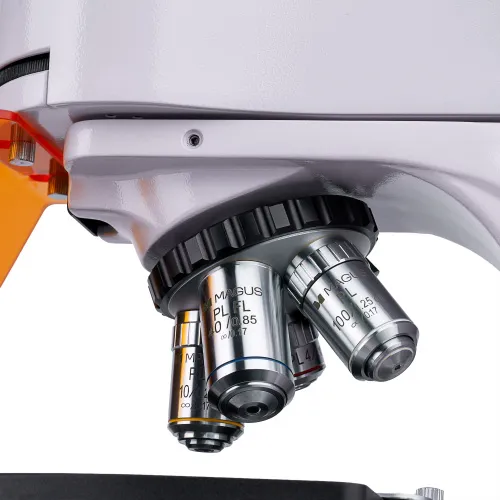
Five objectives can be installed in the revolving eyepiece. Four infinity plan achromatic objectives are included. One of them, a fluo objective with a 40x magnification, is designed for fluorescence observations. 4x, 10x, and 100x objectives are used for the brightfield microscopy technique. An additional objective can be mounted in the free slot of the revolving nosepiece to achieve extra magnification within the magnification range of the microscope.
The revolving nosepiece with objectives is oriented toward the interior – the user can see the objective inserted into the optical path.
Illumination
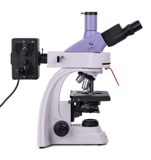
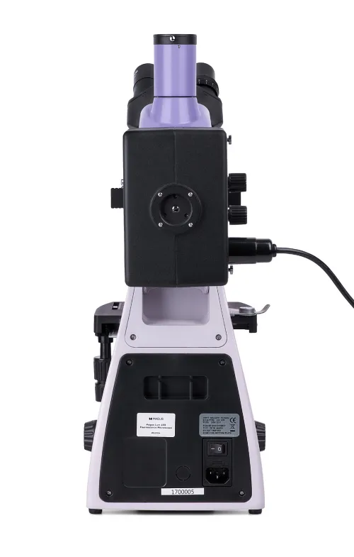
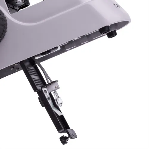
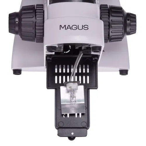
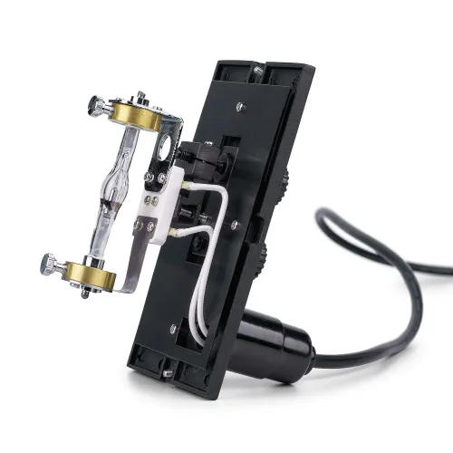
The reflected light source is a 100W mercury lamp. It is placed inside the lamphouse, which provides efficient heat dissipation: The lamp does not overheat even during long hours of operation. The lamp can be centered along three axes to provide the optimal illumination of the working area. If you need to replace it, it will be fairly easy to remove and install the bulb. The mercury lamp is suitable for fluorescent dyes because it has discrete peaks. The reflected light illuminator contains four excitation filters: ultraviolet (UV), violet (V), blue (B), and green (G).
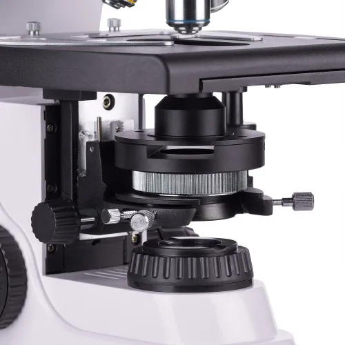
Transmitted light source: 30W halogen bulb. It is bright enough to work with any objectives of any magnification. Thanks to the color temperature of the illumination, even long observations will not strain the eyes. The illumination system also includes a condenser. It has a slot for a slider (phase contrast or darkfield).
In transmitted and reflected light, you can set up the Köhler illumination method. When the illumination is set up in this way, there is no darkening at the edges, the sample is clearly visible, and the image artifacts are removed.
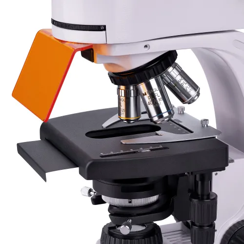
Stage and focusing mechanism
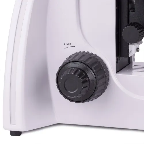
The microscope is equipped with an ergonomically designed stage without a positioning rack. When observing in reflected light, a black stage plate is placed on the stage to remove stray light. The specimen holder is removable and can be taken out, if necessary, by loosening two screws. The focusing knobs are coaxial and located on each side of the microscope. Coarse focusing has a lock knob and tension adjustment and fine focusing is used for focus adjustment.
Accessories
The MAGUS Lum 400 microscope can be equipped with additional accessories: digital camera, phase contrast device, polarization device, darkfield condenser, eyepieces and objectives, darkfield and phase contrast sliders. For measuring objects, the configuration can be supplemented with a calibration slide.
Key features of the microscope:
- Microscopy techniques: fluorescence in reflected light, brightfield in transmitted light
- Other research methods with optional accessories: phase contrast, simple polarization, darkfield
- Trinocular head with a trinocular tube for mounting a digital camera; 360° rotatable eyepiece tubes
- Infinity plan achromatic objectives, including a fluo objective
- Reflected light source: 100W mercury lamp, transmitted light source: 30W halogen lamp
- Köhler illumination method in transmitted and reflected light
- Fluorescence filters: ultraviolet (UV), violet (V), blue (B), green (G)
- Ergonomic stage with a removable specimen holder and no positioning rack
Key features of the camera:
- For fluorescence and darkfield microscopy with 40x, 60x, and 100x objectives
- 2.3MP resolution for getting detailed images at high magnification; low noise level, low-power dissipation
- 120fps for observing moving objects, recording video, and moving the sample without jerkiness or delays
- Global shutter for fast signal readout from the sensor, increased image brightness, and no false optical effects when observing moving objects
- SONY Exmor monochrome CMOS backlit sensor provides low noise level and high light sensitivity even in low-light conditions. You will get clearer, brighter, and more color-saturated images.
- USB3.0 interface for fast, smooth data transfer
- Software with photo, video recording, editing, external display functions, linear and angular measurements
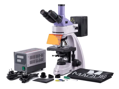
The kit includes:
- MAGUS CLM10 Digital Camera (digital camera, USB cable, installation CD with drivers and software, user manual and warranty card)
- Stand with transmitted light source, focusing mechanism, stage, condenser mount, and revolving nosepiece
- Abbe condenser
- Reflected light illuminator
- Mercury lamphouse
- Trinocular head
- Infinity plan achromatic objective: PL 4x/0.10 WD 19.8mm
- Infinity plan achromatic objective: PL 10x/0.25 WD 5.0mm
- Infinity plan achromatic objective, fluo: PL FL 40x/0.85 (spring-loaded) WD 0.42mm
- Infinity plan achromatic objective: PL 100x/1.25 (spring-loaded, oil) WD 0.36mm
- Eyepiece 10x/22mm with long eye relief (2 pcs.)
- Eyepiece eyecup (2 pcs.)
- UV shield
- Filter slider
- Black protective plate for under-stage installation
- C-mount camera adapter
- Color filter (4 pcs.)
- Bottle of immersion oil
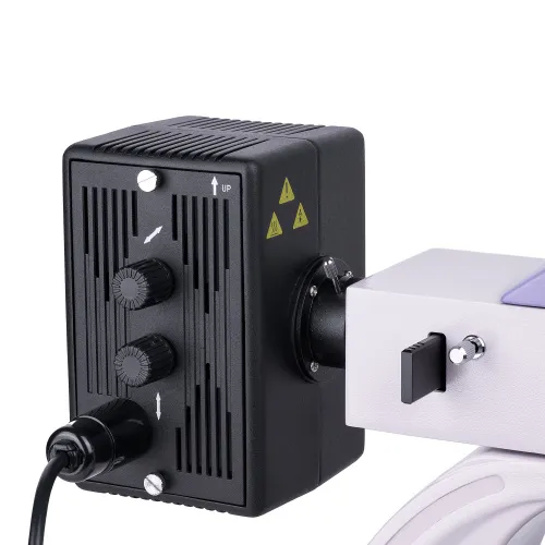
- Mercury lamp power supply
- AC power cord (2 pcs.)Mercury lamphouse power cord
- Dust cover
- User manual and warranty card
Available on request:
- 10x/22mm eyepiece with a scale
- 12.5x/14mm eyepiece (2 pcs.)
- 15x/15mm eyepiece (2 pcs.)
- 20x/12mm eyepiece (2 pcs.)
- 25x/9mm eyepiece (2 pcs.)
- Infinity plan achromatic objective, fluo: PL FL 10x/0.35 WD 2.37mm
- Infinity plan achromatic objective: PL 20х/0.40 WD 8.8mm
- Infinity plan achromatic objective: PL 60x/0.80 ∞/0.17 WD 0.46mm
- Phase contrast device
- Darkfield condenser NA 0.9
- Oil darkfield condenser NA 1.36–1.25
- Darkfield slider
- Polarization device
- Calibration slide
- Monitor and digital camera with an HDMI interface
| Product ID | 83016 |
| Brand | MAGUS |
| Warranty | 5 |
| EAN | 5905555018157 |
| Package size (LxWxH) | 17.7x11.8x37.8 in |
| Shipping Weight | 37.7 lb |
| Type | biological, light/optical, digital |
| Microscope head type | trinocular |
| Head | Gemel head (Siedentopf, 360° rotation) |
| Head inclination angle | 30 ° |
| Magnification, x | 40 — 1000 |
| Magnification, x (optional) | 40–1250/1500/2000/2500 |
| Eyepiece tube diameter, in | 1.2 |
| Eyepieces | 10х/22mm, eye relief: 10mm (*optional: 10x/22mm with scale, 12.5x/14; 15x/15; 20x/12; 25x/9) |
| Objectives | infinity plan achromatic and fluo objectives: PL 4x/0.10, PL 10x/0.25, PL FL 40x/0.85, PL 100x/1.25 (oil); parfocal distance 45mm (*optional: PL FL 10x/0.35, PL 20х/0.40, PL 60x/0.80 ∞/0.17) |
| Revolving nosepiece | for 5 objectives |
| Working distance, mm | 19.8 (4x); 5.0 (10x); 0.42 (FL 40x); 0.36 (100x); 2.37 (FL 10х); 8.8 (20х); 0.46 (60х) |
| Interpupillary distance, in | 1.9 — 3 |
| Stage, mm | 180x150 |
| Stage moving range, mm | 75/50 |
| Stage features | two-axis mechanical stage, without a positioning rack |
| Condenser | Abbe condenser, N.A. 1.25, center-adjustable, height-adjustable, adjustable aperture diaphragm, a slot for a darkfield slider and phase contrast slider, dovetail mount |
| Diaphragm | adjustable aperture diaphragm, adjustable iris field diaphragm |
| Focus | coaxial, coarse focusing (21mm, 39.8mm/circle, with a lock knob and tension adjusting knob) and fine focusing (0.002mm) |
| Body | CNC aluminum alloy |
| Illumination | fluorescent, halogen |
| Brightness adjustment | ✓ |
| Power supply | 85–265V, 50/60Hz (US type plug adapter is not included), AC network |
| Light source type | reflected light: 100W mercury lamp; transmitted light: 12V/30W halogen lamp |
| Light filters | yes |
| Operating temperature range, °F | 41...+95 |
| Ability to connect additional equipment | darkfield slider, phase contrast device (condenser and objectives), darkfield condenser (dry or oil), polarization devices (polarizer and analyzer) |
| User level | experienced users, professionals |
| Assembly and installation difficulty level | complicated |
| Fluorescent module | filters: ultraviolet (UV), violet (V), blue (B), green (G) |
| Fluorescence filter: filter type, excitation wavelength/dichroic mirror/emission wavelength | ultraviolet (UV), 320–380nm/425 nm/435 nm; violet (V), 380–415nm/455nm/475nm; blue (B), 450–490nm/505nm/515nm; green (G), 495–555nm/585nm/595nm |
| Application | laboratory/medical |
| Illumination location | dual |
| Research method | bright field, fluorescence |
| Pouch/case/bag in set | dust cover |
| Camera specifications | |
| Sensor | SONY Exmor CMOS |
| Color/monochrome | monochrome |
| Megapixels | 2.3 |
| Maximum resolution, pix | 1920x1200 |
| Sensor size | 1/1.2" (11.25x7.03 mm) |
| Pixel size, μm | 5.86x5.86 |
| Light sensitivity | 1016mV with 1/30s |
| Signal/noise ratio | 0.15mV at 1/30s |
| Exposure time | 0.244ms–2s |
| Video recording | ✓ |
| Video recording | yes |
| Frame rate, fps at resolution | 120@1920x1200 |
| ADC digit capacity (bit) | 8/12 (selectable) |
| Place of installation | trinocular tube, eyepiece tube instead of an eyepiece |
| Image format | *.jpg, *.bmp, *.png, *.tif |
| Video format | output: *.wmv, *.avi, *.h264 (Windows 8 and later), *h265 (Windows 10 and later) |
| Spectral range, nm | 380–700 (with anti-reflective filter) |
| Shutter type | Global shutter |
| White balance | automatic, manual |
| Exposure control | automatic, manual |
| Software features | brightness, exposure time, image size |
| Software | MAGUS View |
| Output | 5Gb/s, USB 3.0 |
| System requirements | Windows 8/10/11 (32bit and 64bit), Mac OS X, Linux, up to 2.8GHz Intel Core 2 or higher, minimum 2GB RAM, USB 3.0 port, CD-ROM, 17" or larger display |
| Mount type | C-mount |
| Camera power supply | DC, 5V, from computer USB port; a 12V, 3A adapter for Peltier element |
| Camera operating temperature range, °F | -10...+50 |
| Operating humidity range, % | 30 — 80 |
and downloads
We have gathered answers to the most frequently asked questions to help you sort things out
Find out why studying eyes under a microscope is entertaining; how insects’ and arachnids’ eyes differ and what the best way is to observe such an interesting specimen
Read this review to learn how to observe human hair, what different hair looks like under a microscope and what magnification is required for observations
Learn what a numerical aperture is and how to choose a suitable objective lens for your microscope here
Learn what a spider looks like under microscope, when the best time is to take photos of it, how to study it properly at magnification and more interesting facts about observing insects and arachnids
This review for beginner explorers of the micro world introduces you to the optical, illuminating and mechanical parts of a microscope and their functions
Short article about Paramecium caudatum - a microorganism that is interesting to observe through any microscope