BG
BG  BY
BY  CY
CY  CZ
CZ  DE
DE  EE
EE  ES
ES  EU
EU  GR
GR  HU
HU  IS
IS  IT
IT  LT
LT  LV
LV  MY
MY  PL
PL  PT
PT  RO
RO  SK
SK  TR
TR  UA
UA MAGUS Lum VD500 Fluorescence Inverted Digital Microscope
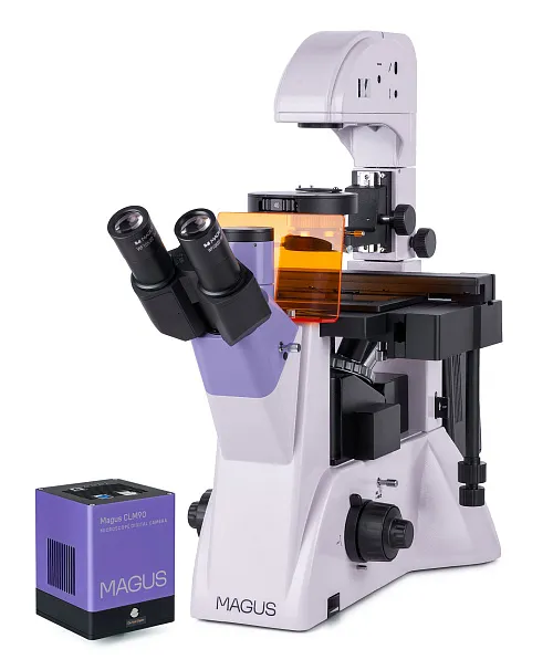
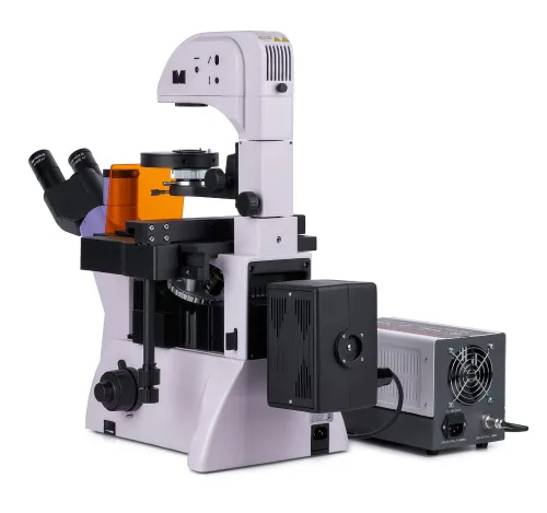
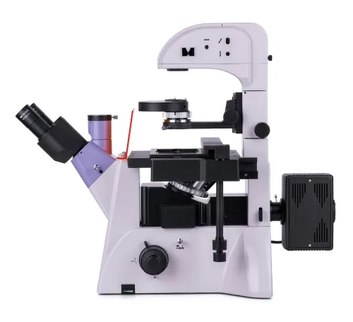


With a camera. Magnification: 100–400x. Trinocular head, plan achromatic objectives, reflected light source – 100W mercury lamp, transmitted light source – 30W halogen bulb, Köhler illumination
| Product ID | 83020 |
| Brand | MAGUS |
| Warranty | 5 years |
| EAN | 5905555018218 |
| Package size (LxWxH) | 15x24x32.3 in |
| Shipping Weight | 63.3 lb |
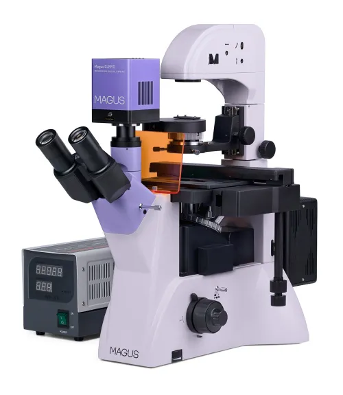
The MAGUS Lum V500 Fluorescent Inverted Microscope is designed for observing samples in laboratory ware up to 55mm high and a bottom thickness of up to 1.2mm. When the illuminator stand is tilted, it is also possible to use larger lab ware of up to 165mm. You can observe samples in the reflected light (fluorescence microscopy technique) and transmitted light (brightfield and phase-contrast microscopy). The main areas of microscope application: medicine, pharmacology, biochemistry, epidemiology, etc.
Digital Camera
The MAGUS CLM90 digital camera is designed for use in fluorescence and darkfield microscopy techniques.
The camera is equipped with a 7.1MP sensor and produces realistic images at a resolution of 3200x2200px. The camera is recommended for use with 4x, 10x, 20x, and 40x objectives. When working with low magnification objectives, the camera will allow you to see the finer details.
Video is recorded at 51.3fps or 133.8fps depending on its resolution. Videos are smooth with soft and subtle transitions between frames. The movement of the sample is displayed in real time with no delays. The camera makes it easy to work with objects that are moving and is suitable for classroom demonstrations.
The camera is equipped with a USB3.0 interface. The data transfer speed is 10 times faster than USB2.0 cameras. The high-speed camera is recommended for professional laboratories, research, or university training.
Optics
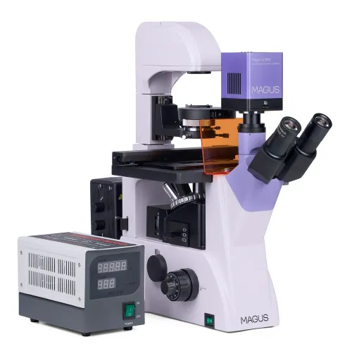
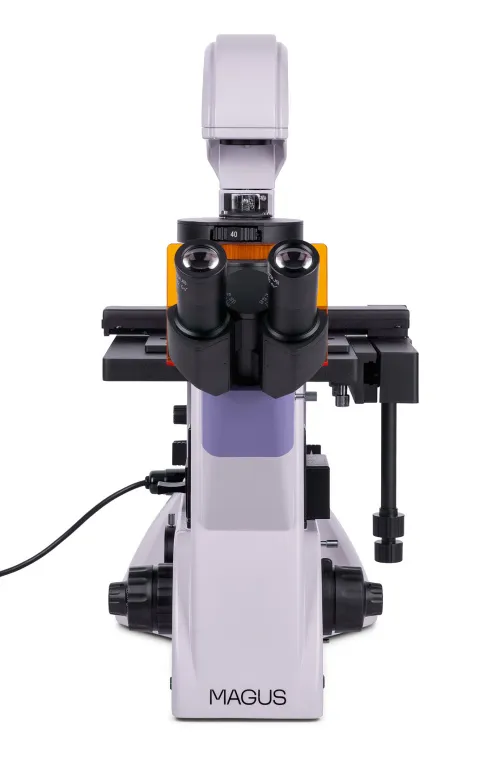
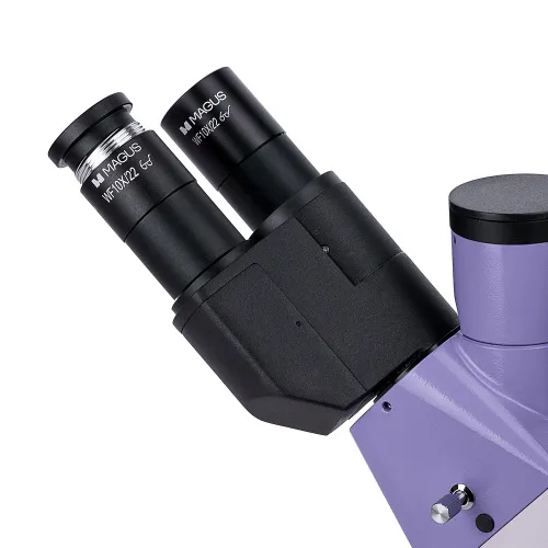
The microscope is equipped with a trinocular head with a trinocular tube. There is a side camera port on the microscope. This design allows you to mount a digital camera and monitor to the microscope at the same time. The beam splitting on the trinocular head is 100/0 or 50/50, on the body – 100/0 or 0/100. The eyepiece tubes are 180° rotatable, which allows you to adjust the eye relief to fit the user.
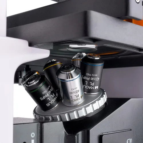
The revolving nosepiece is located on the stand under the stage. It has six free slots for mounting objectives. Three phase contrast objectives as well as three fluo and brightfield objectives are included. There are plan achromatic objectives with a long working distance designed for lab ware with a bottom thickness of 1.2mm.
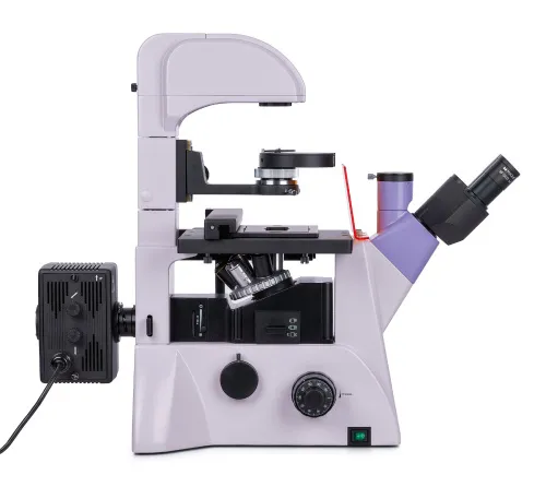
Illumination
The reflected light source is a 100W mercury lamp. It is placed in the lamphouse for better heat dissipation and a lower risk of overheating during long hours of operation. The lamphouse has a wide range of wavelengths and peaks, making it convenient to work with a variety of fluorescent dyes. Fluorescence filters: ultraviolet (UV), violet (V), blue (B), and green (G). The mercury lamp shines brightly, provides good visibility, and can be easily replaced when needed.
The transmitted light is provided by a 30W halogen lamp. It shines brightly and the spectrum of light is warm. Therefore, observing with each objective is efficient and comfortable for the eyes. The microscope has a phase-contrast condenser with four slots: One is used for the brightfield and the other three are for phase-contrast objectives at 10x, 20x, and 40x. Phase rings can be centered. You can switch from brightfield to phase contrast and back again by rotating the disk. It is convenient for research as it saves time during work.
The Köhler illumination method for the transmitted and reflected light. The application of this method allows you to obtain a clear, sharp image in high resolution without artifacts and darkening at the edges.
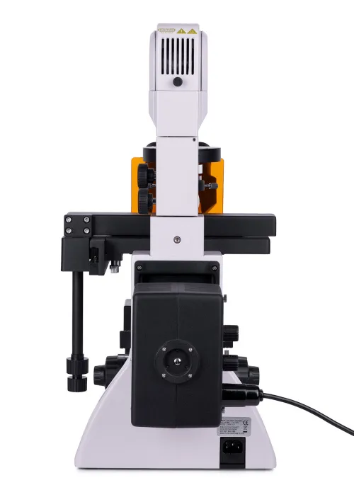
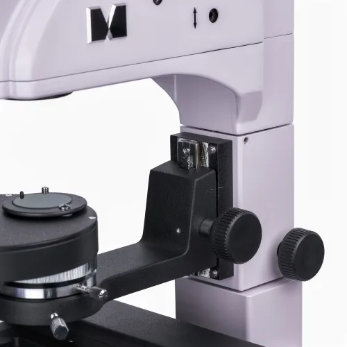
Stage and focusing mechanism
The stage is fixed and has a mechanism for moving lab ware. The lab ware can be moved horizontally in two mutually perpendicular directions. The movement is precise and smooth, and it comes with four dish cup holders to hold the ware of different sizes. The focusing of the microscope is adjusted by coarse and fine focusing knobs. The coarse focusing has a lock knob and tension adjustment. The focusing knobs are located coaxially on both sides of the body.
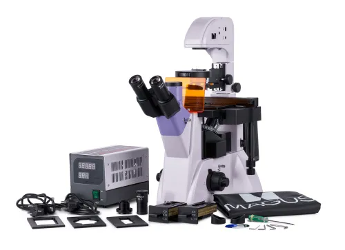
Accessories
The product line includes additional accessories for the MAGUS Lum V500 microscope: eyepieces, digital cameras, and calibration slides.
Key features of the microscope:
- Fluorescence microscopy in the reflected light as well as brightfield and phase contrast microscopy in the transmitted light
- Option to observe samples in laboratory ware up to 55mm as well as up to 165mm when the stand is tilted
- 100W mercury lamp for reflected light, 30W halogen lamp for transmitted light; Köhler illumination setup
- Four fluorescence filters: ultraviolet (UV), violet (V), blue (B), and green (G)
- Trinocular head, 180° rotatable. Two options for mounting a digital camera and monitor: the 100/0 or 50/50 beam splitting (trinocular head) or the 100/0 or 0/100 beam splitting (on the microscope)
- Stage with dish holders for lab ware of different diameters
- Phase-contrast condenser with a brightfield aperture and three phase-contrast apertures
Key features of the camera:
- For fluorescence and darkfield microscopy with 4x, 10x, 20x, or 40x objectives
- 7.1MP resolution: When observing with low magnification objectives, the camera will enable you to see finer details
- 51.3fps and 133.8fps at 3200x2200px and 1584x1100px resolutions for observing moving objects, recording video and moving the preparation without jerkiness and delays
- Peltier element reduces the sensor temperature during prolonged operation and eliminates thermal noise and, therefore, long shutter speeds can be used when taking photos
- Global shutter for fast signal readout from the sensor, increased image brightness, no false optical effects when observing moving objects
- SONY Exmor monochrome CMOS backlit sensor provides low noise level and high light sensitivity even in low-light conditions. You will get clearer, brighter, and more color-saturated images
- USB3.0 interface for fast, smooth data transfer
- Software with photo, video recording, editing, external display functions, linear and angular measurements
The kit includes:
- MAGUS CLM90 Digital Camera (digital camera, USB cable, 12V, 3A adapter, case, Installation CD with drivers and software, user manual and warranty card)
- Base with a power input, transmitted light source and condenser, focusing mechanism, stage, and revolving nosepiece
- Fluorescence units
- Mercury lamphouse
- Trinocular head
- Infinity plan achromatic objective: PLL 10x/0.25 WD 4.3mm
- Infinity plan achromatic objective: PLL 20х/0.40 WD 8.0mm
- Infinity plan achromatic objective: PLL 40х/0.60 WD 3.5mm
- Infinity plan achromatic objective: PLL 10x/0.25 PHP2 WD 4.3mm
- Infinity plan achromatic objective: PLL 20x/0.40 PHP2 WD 8.0mm
- Infinity plan achromatic objective: PLL 40x/0.60 PHP2 WD 3.5mm
- Eyepiece 10x/22mm with long eye relief (2 pcs.)
- UV shield
- C-mount adapter 1x
- Hex key wrench
- Mercury lamphouse power supply
- Power cord
- Reflected light illuminator power cord
- Dust cover
- User manual and warranty card
Available on request:
- 10x/22mm eyepiece with a scale
- 12.5x/14mm eyepiece (2 pcs.)
- 15x/15mm eyepiece (2 pcs.)
- 20x/12mm eyepiece (2 pcs.)
- 25x/9mm eyepiece (2 pcs.)
- Calibration slide
| Product ID | 83020 |
| Brand | MAGUS |
| Warranty | 5 years |
| EAN | 5905555018218 |
| Package size (LxWxH) | 15x24x32.3 in |
| Shipping Weight | 63.3 lb |
| Camera specifications | |
| Type | biological, light/optical |
| Microscope head type | trinocular |
| Head | Siedentopf, rotatable 180° |
| Magnification, x | 100 — 400 |
| Magnification, x (optional) | 40–500/600/800/1000 |
| Eyepiece tube diameter, in | 1.2 |
| Eyepiece tube diameter, in | 1.2 |
| Eyepieces | 10х/22mm, eye relief: 10mm (*optional: 10x/22mm with scale, 12.5x/14; 15x/15; 20x/12; 25x/9) |
| Objectives | infinity plan achromatic: PLL 10x/0.25/4.3; PLL 20x/0.40/8.0; PLL 40x/0.60/3.5; phase: PLL 10x/0,25/4,3 PHP2; PLL 20x/0.40/8.0 PHP2; PLL 40x/0.60/3.5 PHP2; parfocal distance: 45mm |
| Revolving nosepiece | for 6 objectives |
| Working distance, mm | 4.3 (10х); 8.0 (20x); 3.5 (40x) |
| Interpupillary distance, in | 1.9 — 3 |
| Interpupillary distance, in | 1.9 — 3 |
| Stage, mm | 227x208 |
| Stage moving range, mm | 77/134.5 |
| Stage features | fixed, with glass plate Ø118mm and mechanical attachment; dish holders: 86x129.5, Ø90mm; 34x77.5mm, Ø68.5mm; 57x82mm, Ø60mm; 29x77.5mm, Ø35mm |
| Condenser | NA 0.6, working distance: 55mm; phase contrast turret; with locking screws |
| Diaphragm | adjustable aperture diaphragm, adjustable iris field diaphragm |
| Focus | coaxial, coarse (with coarse focusing tension adjustment and a lock knob), and fine (0.002mm) |
| Body | CNC aluminum alloy |
| Illumination | fluorescent, halogen |
| Brightness adjustment | ✓ |
| Power supply | 85–265V, 50/60Hz (US type plug adapter is not included), AC network |
| Light source type | reflected light: 100W mercury lamp; transmitted light: 12V/30W halogen lamp |
| Light filters | yes |
| Operating temperature range, °F | 41...+95 |
| Operating temperature range, °F | 41...+95 |
| User level | experienced users, professionals |
| Assembly and installation difficulty level | complicated |
| phase contrast condenser (turret) with a free slot and phase annuli plates for 10x, 20x and 40x objectives; centering auxiliary microscope | |
| Fluorescent module | filters: ultraviolet (UV), violet (V), blue (B), green (G) |
| Fluorescence filter: filter type, excitation wavelength/dichroic mirror/emission wavelength | ultraviolet (UV), 320–380nm/425 nm/435 nm; violet (V), 380–415nm/455nm/475nm; blue (B), 450–490nm/505nm/515nm; green (G), 495–555nm/585nm/595nm |
| Application | laboratory/medical |
| Illumination location | dual |
| Research method | bright field, fluorescence, phase-contrast microscopy |
| Pouch/case/bag in set | dust cover |
| Sensor | SONY Exmor CMOS |
| Color/monochrome | monochrome |
| Megapixels | 7.1 |
| Maximum resolution, pix | 3200x2200 |
| Sensor size | 1.1" (14.4x9.9mm) |
| Pixel size, μm | 4.5x4.5 |
| Two-stage thermoelectric module (Peltier element) to set the temperature 42°C below room temperature | ✓ |
| Light sensitivity | 3354mV with 1/30s |
| Signal/noise ratio | 0.15mV at 1/30s |
| Exposure time | 0.1ms–1h |
| Video recording | ✓ |
| Frame rate, fps at resolution | 133.8@1584x1100, 51.3@3200x2200 |
| ADC digit capacity (bit) | 8/12 (selectable) |
| Image format | *.jpg, *.bmp, *.png, *.tif |
| Video format | output: *.wmv, *.avi, *.h264 (Windows 8 and later), *h265 (Windows 10 and later) |
| Spectral range, nm | 380–650 (with anti-reflective filter) |
| Shutter type | Global shutter |
| White balance | automatic, manual |
| Exposure control | automatic, manual |
| Software features | brightness, exposure time, image size |
| Software | MAGUS View |
| Output | 5Gb/s, USB 3.0 |
| System requirements | Windows 8/10/11 (32bit and 64bit), Mac OS X, Linux, up to 2.8GHz Intel Core 2 or higher, minimum 2GB RAM, USB 3.0 port, CD-ROM, 17" or larger display |
| Mount type | C-mount |
| Camera power supply | DC, 5V, from computer USB port; a 12V, 3A adapter for Peltier element |
| Camera operating temperature range, °F | -10...+50 |
| Operating humidity range, % | 30 — 80 |
and downloads
We have gathered answers to the most frequently asked questions to help you sort things out
Find out why studying eyes under a microscope is entertaining; how insects’ and arachnids’ eyes differ and what the best way is to observe such an interesting specimen
Read this review to learn how to observe human hair, what different hair looks like under a microscope and what magnification is required for observations
Learn what a numerical aperture is and how to choose a suitable objective lens for your microscope here
Learn what a spider looks like under microscope, when the best time is to take photos of it, how to study it properly at magnification and more interesting facts about observing insects and arachnids
This review for beginner explorers of the micro world introduces you to the optical, illuminating and mechanical parts of a microscope and their functions
Short article about Paramecium caudatum - a microorganism that is interesting to observe through any microscope