BG
BG  BY
BY  CY
CY  CZ
CZ  DE
DE  EE
EE  ES
ES  EU
EU  GR
GR  HU
HU  IS
IS  IT
IT  LT
LT  LV
LV  MY
MY  PL
PL  PT
PT  RO
RO  SK
SK  TR
TR  UA
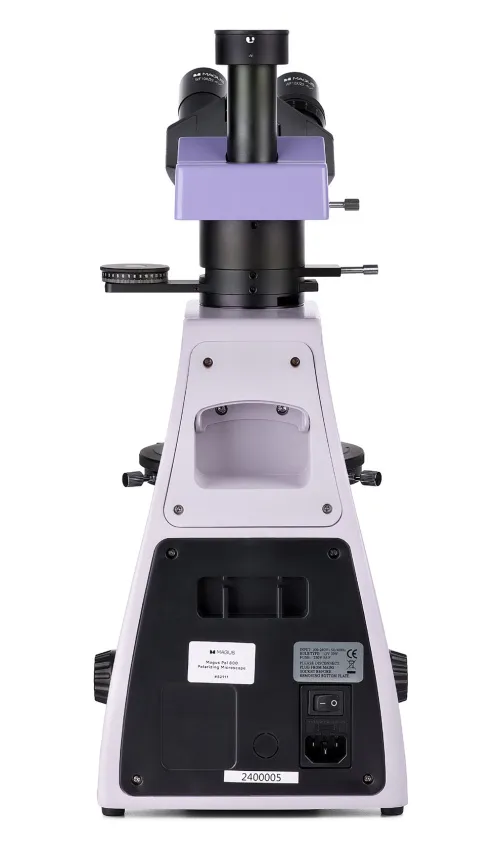
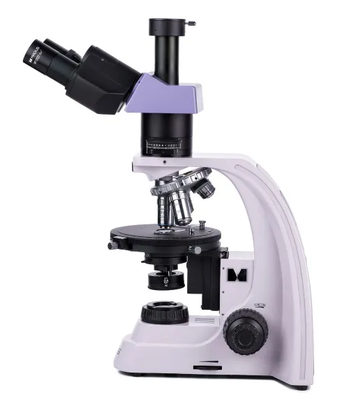
UA MAGUS Pol D800 Polarizing Digital Microscope
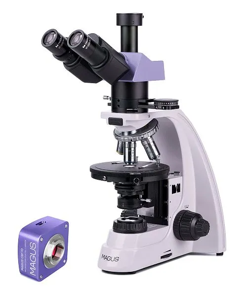
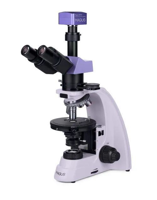
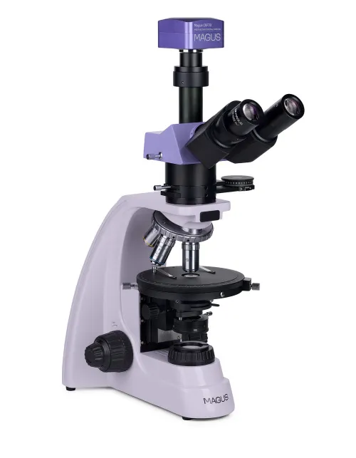

With a camera. Magnification: 40–600x. Trinocular head, plan achromatic objectives. Transmitted light illumination, light source – 30W halogen bulb, Köhler illumination. Bertrand lens and compensators
| Product ID | 83040 |
| Brand | MAGUS |
| Warranty | 5 |
| EAN | 5905555018515 |
| Package size (LxWxH) | 16.9x10.6x24.8 in |
| Shipping Weight | 33.3 lb |
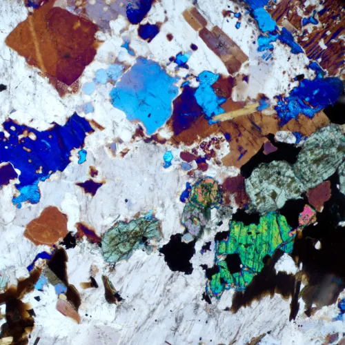
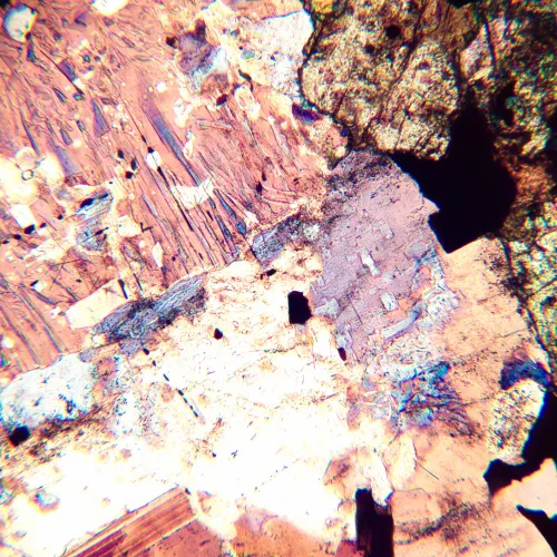
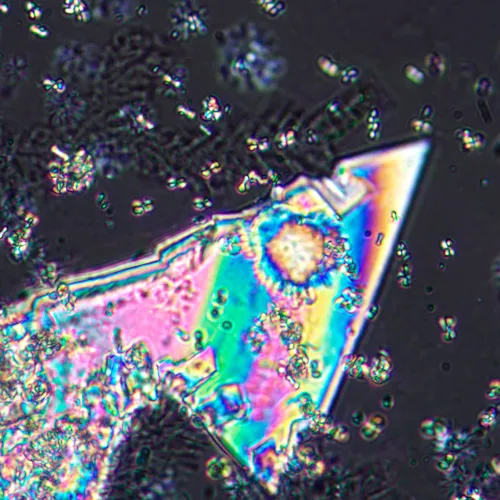
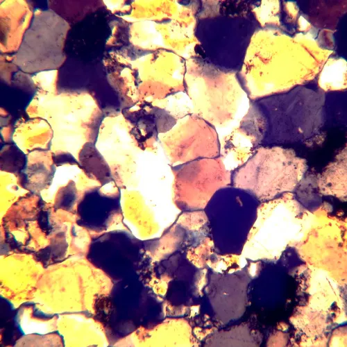
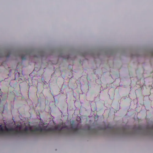
The MAGUS Pol 800 Polarizing Microscope is used to study anisotropic objects in polarized and ordinary transmitted light. Objects of study can be biological, geological, or polymer. The optics of the microscope are strain-free, i.e., they do not create parasitic refractions. They form a clear, high-contrast and authentic image. The microscope’s functionality is useful in various fields of science: medicine, forensics, mineralogy, petrography, crystallography, etc.
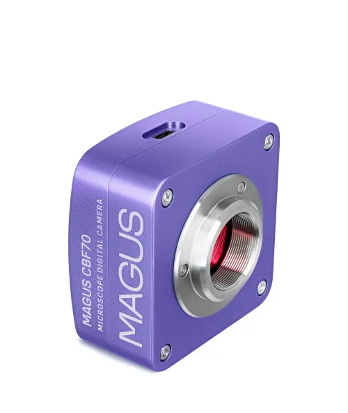
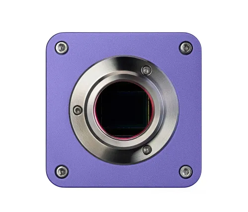
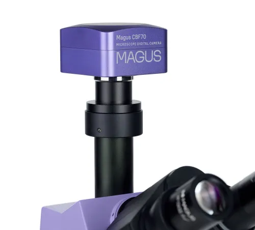
Digital camera
The professional scientific camera MAGUS CBF70 is designed for use with a microscope as well as 4x, 10x, 20x, and 40x objectives. The device enables you to take photographs, shoot videos, organize demonstrations, display images on an external screen, and make angular and linear measurements of objects under study (for this function to work correctly, you must calibrate the software). The camera is recommended for working in the brightfield method.
The MAGUS CBF70 camera has five image resolution and frame rate modes, and so you can easily find among them the one that best suits your needs: 5280x3954px (17fps), 3952x3952px (17fps), 2640x1976px (56fps), 1760x1316px (67fps), and 584x438px (192fps).
High-resolution modes are ideal for photography, enabling you to create clear, vibrant, and highly detailed images. Modes with an increased frame rate will help when recording smooth and informative videos, without jumps and delays, recording the slightest movements of the object or features of observed fast-flowing processes.
To obtain the best image quality without distortions, it is recommended to use an adapter with 1x magnification.
The camera design uses a SONY Exmor 4/3'' color backlit CMOS sensor. It makes the image bright even in low ambient light. The larger physical size of the sensor helps the colors displayed appear deeper and the picture clearer.
Optics
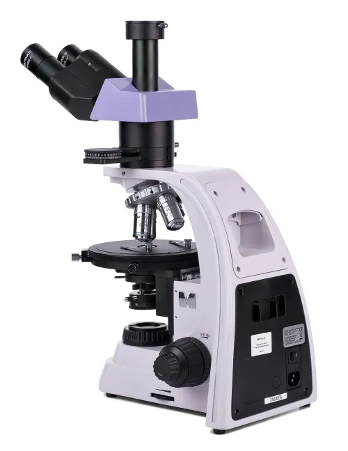
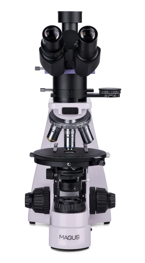
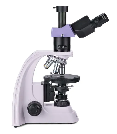
The trinocular head is equipped with a trinocular tube for mounting a digital camera. By using the switch on the body, the light is completely directed either to the eyepiece tubes or to the digital camera. The 30° angle of the eyepiece tubes is comfortable for long-term observations and does not cause strain on the neck muscles. The left tube has a diopter adjustment ring in which it rotates and adapts the optics of the microscope to the user’s unique vision.
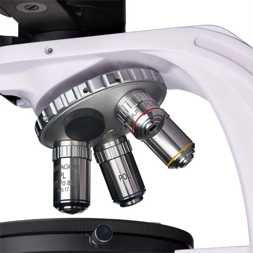
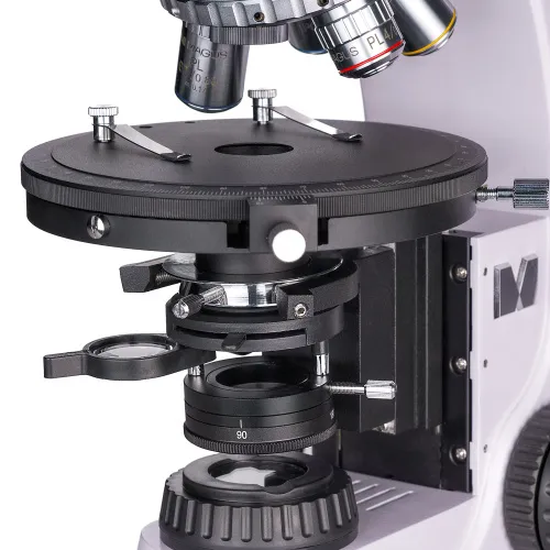
The revolving nosepiece is designed for the simultaneous installation of five objectives, with four already included in the package. An additional objective from the MAGUS range of accessories can be purchased separately. All of the objectives are designed to work with 0.17mm thick cover slips. Thanks to the “away from the observer” revolver design, the objective inserted into the optical path is clearly visible during work, which makes the researcher’s work more convenient. The revolver nosepiece slots are centered.
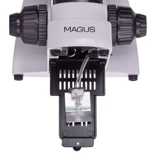
Illumination
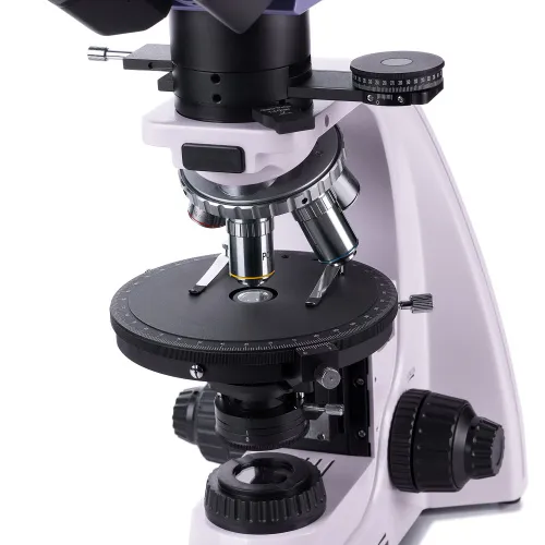
The illumination source is located under the object stage, i.e., observations are carried out in transmitted light. The 30W halogen bulb produces bright, eye-friendly illumination that is suitable for use on all objectives. Adjusting the illumination using the Köhler method achieves higher contrast and enhanced image quality.
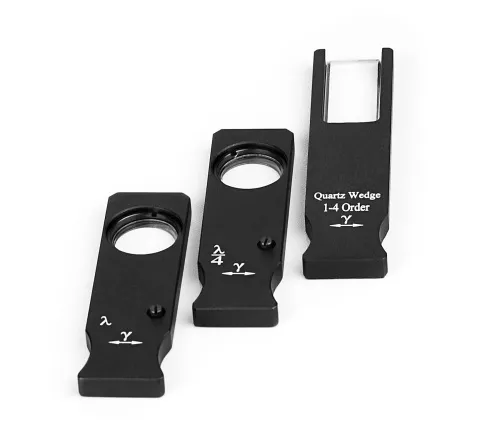
The microscope is equipped with a polarizer and analyzer. To work in polarized light, the analyzer is introduced into the optical path, and the polarization angle is changed by rotating the polarizer and analyzer relative to each other. The microscope also has an intermediate attachment that holds a Bertrand lens and has a slot for compensators. Compensators enhance the contrast of samples with low birefringence, and the Bertrand lens is designed for conoscopic studies.
Stage and focusing mechanism
The microscope stage rotates and that enables you to quickly change the refraction of light by the sample when working in polarized light. The stage is centered relative to the optical axis of the microscope, has rotation angle gradation and a scale that enables you to determine the angle with an accuracy of 0.1°.
The focusing mechanism provides coarse and fine sharpness adjustment. For coarse focusing, a coarse focusing lock is available, and you can also adjust the coarse tension for the most comfortable control.
Accessories
Optional eyepieces, objectives, digital cameras, and calibration slides are available for the microscope. They broaden the capabilities of the microscope for carrying out researchers’ tasks.
Key features of the microscope:
- Observations in transmitted light in a brightfield and by the polarization methods
- Illumination source is a 30W halogen bulb with a “warm” spectrum of light that is comfortable for the eyes
- Polarization is achieved by a rotating analyzer and polarizer; the intermediate attachment has a Bertrand lens and a slot for compensators
- It is possible to adjust illumination according to the Köhler method, which improves image readability
- The trinocular tube allows for the installation of a digital camera in the trinocular tube
- The infinity plan achromatic objectives are strain-free; the revolving nosepiece slots are centered
- Rotating stage, coarse, and fine focusing
- Compatible with additional accessories: eyepieces, objectives, etc.
Key features of the camera:
- Five graphics and fps settings: Among them, you can easily find the most suitable mode for you
- Fast data transfer thanks to the USB3.0 interface
- Large SONY Exmor color sensor for bright, clear images even in low light conditions
- “Quick start”: Install the included software and then the camera is ready to use
- Simplify your observations or demonstrate to an audience by displaying your camera image on an external screen
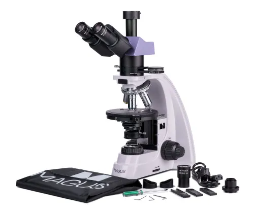
The kit includes:
- MAGUS CBF70 Digital Camera (digital camera, USB cable, installation CD with drivers and software, user manual and warranty card)
- Base with a power input, transmitted light source and condenser, focusing mechanism, stage, and revolving nosepiece
- Trinocular head
- Intermediate tube with Bertrand lens, an analyzer, and a compensator slot
- Compensators: λ compensator, λ/4 compensator, quartz wedge
- Infinity plan achromatic objective: PL 4x/0.10 WD 21mm
- Infinity plan achromatic objective: PL 10x/0.25 WD 5.0mm
- Infinity plan achromatic objective: PL 40x/0.65 (spring-loaded) WD 0.66mm
- Infinity plan achromatic objective: PL 60x/0.80 (spring-loaded) WD 0.46mm
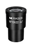
- Eyepiece 10x/22mm with long eye relief (2 pcs.)
- 10x/22mm eyepiece with a scale. Scale value: 0.1mm
- C-mount adapter 1x
- Hex key wrench
- Power cord
- Dust cover
- User manual and warranty card
Available on request:
- Eyepiece 10x/22mm with crosshairs
- 12.5x/14mm eyepiece (2 pcs.)
- 15x/15mm eyepiece (2 pcs.)
- 20x/12mm eyepiece (2 pcs.)
- 25x/9mm eyepiece (2 pcs.)
- Infinity plan achromatic objective: PL 5x/0.12 WD 26.1mm
- Infinity plan achromatic objective: PL 20x/0.4 WD 8.8mm
- Infinity plan achromatic objective, fluo: PL 100x/1.25 (spring-loaded, oil) WD 0.36mm
- Infinity plan achromatic objective, fluo: PL 100x/0.8 (spring-loaded, dry) WD 2.02mm
- Mechanical stage attachment
- Calibration slide
| Product ID | 83040 |
| Brand | MAGUS |
| Warranty | 5 |
| EAN | 5905555018515 |
| Package size (LxWxH) | 16.9x10.6x24.8 in |
| Shipping Weight | 33.3 lb |
| Type | biological, light/optical, digital |
| Microscope head type | trinocular |
| Head | Siedentopf |
| Head inclination angle | 30 ° |
| Magnification, x | 40 — 600 |
| Eyepiece tube diameter, in | 1.2 |
| Eyepieces | 10х/22mm, eye relief: 10mm; 10х/22mm with a scale (*optional: 10x/22mm with crosshairs, 12.5x/14; 15x/15; 20x/12; 25x/9) |
| Objectives | infinity plan achromatic, strain-free: PL 4x/0.10; PL 10x/0.25; PL 40x/0.65; PL 60x/0.80 (*optional: PL 5x/0.12; PL 20x/0.4; PL 100x/1.25 (oil); PL 100x/0.8 (dry)); parfocal height 45mm, designed for work with a 0.17mm thick cover slips |
| Revolving nosepiece | for 5 objectives, centering |
| Working distance, mm | 21 (4x); 5.0 (10x); 0.66 (40x); 0.46 (60x); 26.1 (5x); 8.8 (20x); 0.36 (100x (oil)); 2.02 (100x (dry)) |
| Interpupillary distance, in | 1.9 — 3 |
| Stage, mm | Ø150 |
| Stage features | 360 ° rotatable, centering, 1° graduation of the rotation angle; a vernier scale for measuring angles with an accuracy of 0.1° |
| Eyepiece diopter adjustment, diopters | ±5 (on the left tube) |
| Condenser | Abbe condenser NA 1.25 with an adjustable aperture diaphragm and a swivel lens |
| Diaphragm | adjustable aperture diaphragm, adjustable iris field diaphragm |
| Focus | coaxial, coarse focusing (21mm, 39.8mm/circle, with a lock knob and tension adjusting knob) and fine focusing (0.002mm) |
| Body | solid aluminum |
| Illumination | halogen |
| Brightness adjustment | ✓ |
| Power supply | 220±22V, 50Hz, AC network |
| Light source type | halogen lamp: 12V/30W |
| Operating temperature range, °F | 41...+95 |
| User level | experienced users, professionals |
| Assembly and installation difficulty level | complicated |
| Polarizer | with 0°, 90°, 180°, 270° marks on the scale; 360° rotatable |
| Intermediate tube | built-in analyzer with rotation 0–360°; a vernier scale for measuring angles with an accuracy of 0.1°; Bertrand lens; slot for installing compensators |
| Compensator | λ compensator; λ/4 compensator; quartz wedge |
| Application | laboratory/medical |
| Illumination location | lower |
| Research method | bright field, polarization |
| Pouch/case/bag in set | dust cover |
| Camera specifications | |
| Sensor | SONY Exmor CMOS |
| Color/monochrome | color |
| Megapixels | 21 |
| Maximum resolution, pix | 5280x3956 |
| Sensor size | 4/3" (17.4x13.0mm) |
| Pixel size, μm | 3.3x3.3 |
| Exposure time | 0.1ms–15s |
| Video recording | ✓ |
| Video recording | yes |
| Active range, dB | 72 |
| Place of installation | trinocular tube, eyepiece tube instead of an eyepiece |
| Image format | *.jpg, *.bmp, *.png, *.tif |
| Video format | output: *.wmv, *.avi, *.h264 (Windows 8 and later), *h265 (Windows 10 and later) |
| Spectral range, nm | 380–650 (built-in IR filter) |
| Shutter type | ERS (electronic rolling shutter) |
| White balance | automatic, manual |
| Exposure control | automatic, manual |
| Software features | brightness, exposure time, image size |
| Software | MAGUS View |
| Mount type | C-mount |
| Camera power supply | DC, 5V, from computer USB port; a 12V, 3A adapter for Peltier element |
| Camera operating temperature range, °F | -10...+50 |
| Operating humidity range, % | 20 — 80 |
and downloads
We have gathered answers to the most frequently asked questions to help you sort things out
Find out why studying eyes under a microscope is entertaining; how insects’ and arachnids’ eyes differ and what the best way is to observe such an interesting specimen
Read this review to learn how to observe human hair, what different hair looks like under a microscope and what magnification is required for observations
Learn what a numerical aperture is and how to choose a suitable objective lens for your microscope here
Learn what a spider looks like under microscope, when the best time is to take photos of it, how to study it properly at magnification and more interesting facts about observing insects and arachnids
This review for beginner explorers of the micro world introduces you to the optical, illuminating and mechanical parts of a microscope and their functions
Short article about Paramecium caudatum - a microorganism that is interesting to observe through any microscope